Yellow mucus often triggers concern among patients, yet its presence represents a complex physiological response that requires careful clinical interpretation. The colour transformation from clear to yellow secretions signals specific immune system activities and cellular responses occurring within the respiratory tract. Understanding the underlying mechanisms behind yellow mucus formation helps distinguish between normal immune responses and pathological conditions requiring medical intervention.
The significance of yellow mucus extends beyond mere colour observation, encompassing intricate biochemical processes involving neutrophil activation, enzymatic reactions, and inflammatory cascades. Modern respiratory medicine recognises yellow sputum as a valuable diagnostic indicator, though its interpretation must consider patient history, symptom duration, and accompanying clinical manifestations. This comprehensive examination of yellow mucus explores its formation mechanisms, associated pathogens, diagnostic implications, and therapeutic considerations across various respiratory conditions.
Mucus colour classification and clinical significance in respiratory health
Respiratory mucus serves as the body’s first line of defence against inhaled pathogens, particulates, and environmental irritants. The mucociliary escalator system continuously produces approximately 1.5 litres of mucus daily, maintaining optimal respiratory tract hydration and facilitating pathogen clearance. Under normal physiological conditions, this protective secretion remains virtually invisible, allowing unimpeded breathing whilst performing crucial defensive functions.
The colour spectrum of respiratory secretions provides valuable clinical information about underlying pathological processes. Clear mucus typically indicates normal physiological function or mild irritation from allergens or environmental factors. However, colour changes reflect specific cellular responses and biochemical alterations occurring within the respiratory epithelium and surrounding tissues.
Clear to white mucus production during normal respiratory function
Clear mucus represents the baseline secretion produced by goblet cells and submucosal glands throughout the respiratory tract. This transparent fluid contains water, mucins, proteins, electrolytes, and antimicrobial peptides that collectively maintain respiratory health. The consistency remains thin and easily expectorated, facilitating normal mucociliary clearance mechanisms.
White mucus typically emerges during early viral infections or mild inflammatory responses. The opacity results from increased cellular content, including epithelial cells, inflammatory mediators, and protein concentrations. This colour change often precedes more significant inflammatory responses, serving as an early indicator of immune system activation.
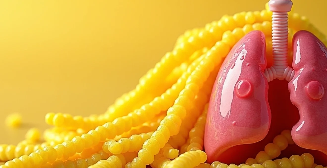
Yellow mucus formation through neutrophil infiltration and myeloperoxidase activity
Yellow mucus development involves complex immunological processes centred around neutrophil recruitment and activation. When respiratory tissues encounter bacterial pathogens or persistent irritants, chemotactic signals attract neutrophils to infection sites. These specialised white blood cells migrate from blood vessels into respiratory tissues, where they engage in active pathogen elimination.
The characteristic yellow colouration primarily results from myeloperoxidase , a haem-containing enzyme abundant in neutrophil granules. During degranulation processes, myeloperoxidase releases into surrounding tissues, creating the distinctive yellow-green pigmentation associated with purulent secretions. This enzymatic activity generates hypochlorous acid, contributing to antimicrobial activity whilst simultaneously producing the colour change clinicians recognise as indicative of bacterial involvement.
Green mucus development via bacterial infection and oxidative stress markers
Green mucus represents an intensification of the inflammatory response observed in yellow secretions. The progression from yellow to green typically indicates increased neutrophil activity, prolonged inflammatory responses, or more virulent bacterial infections. Verdoperoxidase activity and chlorophyll-like compounds contribute to this deeper pigmentation, suggesting sustained immune system engagement.
The green colouration often correlates with bacterial biofilm formation and chronic inflammatory conditions. Pseudomonas aeruginosa infections particularly produce distinctive green sputum due to pyocyanin and pyoverdine pigment production. This colour change frequently accompanies increased sputum viscosity, reduced clearance efficiency, and more persistent respiratory symptoms requiring targeted antibiotic therapy.
Brown and red mucus variants indicating haemoglobin presence and tissue damage
Brown and red mucus variants indicate blood presence within respiratory secretions, ranging from fresh haemoglobin to oxidised iron compounds. Fresh blood produces bright red colouration, whilst older blood undergoes oxidation processes creating brown or rust-coloured secretions. These colour changes suggest mucosal damage, inflammation severity, or underlying pathological conditions affecting respiratory tissue integrity.
Pink or rust-coloured sputum often appears in pneumonia cases, particularly those involving Streptococcus pneumoniae infections. The presence of blood products indicates capillary damage, increased vascular permeability, or direct tissue invasion by pathogenic organisms.
Clinical assessment of bloody sputum requires immediate medical evaluation to exclude serious underlying conditions such as lung cancer, tuberculosis, or severe pneumonia.
Bacterial pathogens associated with yellow sputum production
Yellow sputum production correlates strongly with specific bacterial pathogens that commonly colonise respiratory tissues. These microorganisms trigger characteristic inflammatory responses resulting in neutrophil recruitment, enzyme release, and subsequent colour changes in respiratory secretions. Understanding pathogen-specific characteristics helps guide appropriate therapeutic interventions and predict clinical outcomes.
The bacterial species most frequently associated with yellow mucus production include both community-acquired and healthcare-associated pathogens. Each organism demonstrates unique virulence factors, tissue tropism, and inflammatory response patterns that influence clinical presentation and treatment requirements. Antibiotic susceptibility patterns vary significantly among these pathogens, necessitating targeted therapeutic approaches based on local resistance patterns and patient-specific factors.
Streptococcus pneumoniae infections and purulent discharge characteristics
Streptococcus pneumoniae represents the most common bacterial cause of community-acquired pneumonia, frequently producing characteristic yellow-green sputum. This gram-positive diplococcus possesses a polysaccharide capsule that enhances virulence and tissue invasion capabilities. The organism’s ability to evade initial immune responses allows establishment of lower respiratory tract infections with distinctive clinical presentations.
Pneumococcal infections typically produce purulent sputum containing high neutrophil concentrations, cellular debris, and bacterial components. The inflammatory response generates significant myeloperoxidase release, creating the characteristic yellow colouration observed in pneumococcal pneumonia. Rust-coloured sputum may appear in severe cases due to red blood cell presence and haemoglobin breakdown products.
Haemophilus influenzae colonisation and inflammatory response patterns
Haemophilus influenzae frequently colonises respiratory tissues in patients with chronic obstructive pulmonary disease and other chronic respiratory conditions. This gram-negative coccobacillus produces less dramatic colour changes compared to pneumococcal infections but consistently generates yellow mucus through sustained neutrophilic inflammation.
The organism’s lipopolysaccharide components trigger robust innate immune responses, resulting in prolonged neutrophil activation and enzyme release. Chronic H. influenzae colonisation often produces persistent yellow sputum production, reduced lung function, and recurrent exacerbations in susceptible patients. Biofilm formation contributes to antibiotic resistance and chronic inflammatory states characteristic of this pathogen.
Moraxella catarrhalis proliferation in chronic obstructive airways
Moraxella catarrhalis emerges as an increasingly recognised pathogen in chronic respiratory conditions, particularly chronic obstructive pulmonary disease exacerbations. This gram-negative diplococcus produces beta-lactamase enzymes that confer resistance to penicillin-based antibiotics, complicating treatment protocols.
M. catarrhalis infections typically produce moderate yellow sputum changes accompanied by increased cough frequency and dyspnoea. The organism’s ability to adhere to respiratory epithelium and form biofilms contributes to persistent colonisation and recurrent symptoms. Complement resistance mechanisms allow this pathogen to evade certain immune responses, resulting in chronic inflammatory states and sustained mucus production changes.
Staphylococcus aureus secondary infections following viral respiratory illness
Staphylococcus aureus represents a significant cause of secondary bacterial pneumonia, particularly following influenza and other viral respiratory infections. This gram-positive coccus produces numerous virulence factors that enable tissue invasion, immune evasion, and severe inflammatory responses resulting in distinctive sputum characteristics.
Staphylococcal pneumonia often produces thick, purulent yellow sputum with high bacterial loads and extensive neutrophil infiltration. The organism’s production of toxins and enzymes creates substantial tissue damage, contributing to bloody or rust-coloured secretions in severe cases.
Methicillin-resistant Staphylococcus aureus infections require specific antibiotic protocols and careful monitoring due to limited therapeutic options and potential complications.
Inflammatory biomarkers and cellular components in yellow mucus
Yellow mucus contains numerous inflammatory biomarkers and cellular components that provide insights into underlying pathological processes. These molecular signatures help clinicians assess infection severity, monitor treatment responses, and predict clinical outcomes. Advanced laboratory techniques can quantify specific inflammatory mediators, enabling precision medicine approaches to respiratory infections.
The cellular composition of yellow sputum reflects active immune responses occurring within respiratory tissues. Neutrophils predominate in bacterial infections, whilst eosinophils increase in allergic conditions and certain parasitic infections. Macrophage populations vary depending on infection chronicity and tissue repair processes, providing additional diagnostic information about disease progression and resolution.
Neutrophil elastase concentration and proteolytic enzyme activity
Neutrophil elastase represents a key inflammatory mediator present in high concentrations within yellow mucus. This serine protease degrades elastin, collagen, and other structural proteins, contributing to tissue damage whilst facilitating neutrophil migration through tissue barriers. Elevated elastase levels correlate with infection severity and tissue destruction extent.
Proteolytic enzyme activity measurements provide quantitative assessments of inflammatory intensity and neutrophil activation levels. These enzymes contribute to mucus viscosity changes, impaired mucociliary clearance, and tissue remodelling processes observed in chronic respiratory conditions. Alpha-1 antitrypsin deficiency exacerbates elastase-mediated tissue damage, highlighting the importance of protease-antiprotease balance in respiratory health.
Interleukin-8 levels and chemotactic factor expression
Interleukin-8 serves as the primary chemokine responsible for neutrophil recruitment to infection sites. This inflammatory mediator attracts neutrophils from circulation into respiratory tissues, initiating the cascade of events leading to yellow mucus production. IL-8 concentrations correlate with infection severity and neutrophil infiltration extent.
Elevated IL-8 levels in respiratory secretions indicate active inflammatory processes requiring medical attention. This cytokine also promotes angiogenesis and tissue repair mechanisms, suggesting dual roles in pathology and healing. Therapeutic modulation of IL-8 activity represents a potential target for reducing excessive inflammatory responses whilst preserving protective immune functions.
C-reactive protein correlation with systemic inflammatory response
C-reactive protein levels in sputum reflect systemic inflammatory responses accompanying respiratory infections. This acute-phase reactant increases rapidly during bacterial infections, providing valuable diagnostic and prognostic information. CRP measurements help distinguish bacterial from viral infections and monitor treatment efficacy.
Local CRP production within respiratory tissues contributes to innate immune responses through complement activation and opsonisation processes. Elevated sputum CRP levels often precede systemic elevation, suggesting potential utility as an early infection marker. Point-of-care testing enables rapid CRP assessment, facilitating timely therapeutic decisions in acute care settings.
Lactoferrin and lysozyme antimicrobial protein detection
Lactoferrin and lysozyme represent important antimicrobial proteins present in respiratory secretions. These innate immunity components provide first-line defence against bacterial pathogens through iron sequestration and cell wall disruption mechanisms. Their concentrations increase during infections, contributing to yellow mucus protein content.
Lactoferrin demonstrates broad-spectrum antimicrobial activity against bacteria, viruses, and fungi whilst modulating inflammatory responses. Lysozyme specifically targets bacterial cell walls, particularly gram-positive species. Therapeutic supplementation of these proteins shows promise for enhancing natural antimicrobial defences in immunocompromised patients or chronic respiratory conditions.
Diagnostic criteria and laboratory analysis for yellow sputum assessment
Comprehensive yellow sputum assessment requires systematic laboratory analysis combining microscopic examination, bacterial culture, and molecular diagnostics. Standard protocols begin with macroscopic evaluation of colour, consistency, and volume, followed by microscopic analysis to quantify cellular components and identify potential pathogens. Modern diagnostic approaches incorporate rapid molecular techniques that provide results within hours rather than days.
Gram staining remains fundamental for initial bacterial identification, revealing organism morphology and staining characteristics that guide empirical antibiotic selection. The presence of greater than 25 neutrophils and fewer than 10 epithelial cells per low-power field indicates adequate specimen quality for bacterial culture. Quantitative bacterial cultures help distinguish colonisation from active infection, with concentrations exceeding 10^6 colony-forming units per millilitre suggesting clinically significant infection.
Advanced diagnostic techniques include polymerase chain reaction amplification for specific pathogen detection, antimicrobial susceptibility testing, and biomarker quantification. These methods provide rapid, accurate results that enable targeted therapy selection and resistance pattern identification. Inflammatory marker analysis, including procalcitonin and various cytokines, helps assess infection severity and guide treatment duration decisions.
Quality assurance measures ensure reliable diagnostic results through proper specimen collection, transport, and processing protocols. Induced sputum collection techniques may be necessary for patients unable to produce adequate spontaneous specimens.
Contamination prevention and rapid laboratory processing significantly impact diagnostic accuracy and clinical utility of sputum analysis results.
Treatment protocols and antibiotic resistance considerations for yellow mucus infections
Treatment approaches for yellow mucus-associated infections require careful consideration of likely pathogens, local resistance patterns, and patient-specific factors. Empirical antibiotic therapy typically targets common respiratory pathogens whilst awaiting culture results and susceptibility testing. The emergence of antimicrobial resistance necessitates judicious antibiotic use and implementation of antimicrobial stewardship principles.
First-line treatments for community-acquired pneumonia often include beta-lactam antibiotics such as amoxicillin-clavulanate or cephalosporins, depending on severity and risk factors. Macrolide antibiotics provide coverage against atypical pathogens that may not produce typical yellow sputum changes. Fluoroquinolones are reserved for patients with specific risk factors or treatment failures due to resistance concerns.
Chronic obstructive pulmonary disease exacerbations with yellow sputum production typically respond to shorter antibiotic courses targeting Haemophilus influenzae, Moraxella catarrhalis, and Streptococcus pneumoniae. Treatment duration varies from five to seven days for uncomplicated cases, with longer courses reserved for severe exacerbations or patients with frequent recurrences.
Antimicrobial resistance monitoring remains crucial for maintaining treatment efficacy and preventing further resistance development. Healthcare-associated pneumonia requires broader spectrum coverage including Pseudomonas aeruginosa and methicillin-resistant Staphylococcus aureus considerations. Combination therapy may be necessary for severe infections or patients with multiple resistance risk factors, requiring careful monitoring for drug interactions and adverse effects.
Chronic respiratory conditions manifesting yellow mucus production patterns
Several chronic respiratory conditions demonstrate characteristic yellow mucus production patterns that require ongoing management and monitoring. Chronic obstructive pulmonary disease patients frequently experience periodic yellow sputum production during acute exacerbations, often triggered by bacterial superinfection of chronically inflamed airways. These episodes require prompt recognition and treatment to prevent progression and reduce hospitalisation risk.
Bronchiectasis presents with persistent yellow or green sputum production due to chronic bacterial colonisation and impaired mucociliary clearance. Patients typically exhibit daily productive coughs with purulent secretions containing high bacterial loads and inflammatory mediators. Airway clearance techniques and long-term antibiotic strategies help manage symptoms and reduce exacerbation frequency in this challenging condition.
Cystic fibrosis demonstrates progressive yellow mucus production changes related to chronic Pseudomonas aerugin
osa colonisation and other bacterial pathogens. This genetic condition affects chloride transport across epithelial membranes, resulting in thick, viscous secretions that impair normal clearance mechanisms. Recurrent pulmonary exacerbations characterised by yellow-green sputum production require aggressive antibiotic therapy and supportive care to maintain lung function and prevent irreversible damage.Asthma patients may occasionally produce yellow mucus during severe exacerbations, particularly when bacterial superinfection complicates allergic inflammation. The underlying eosinophilic inflammation creates favourable conditions for bacterial colonisation, resulting in mixed inflammatory patterns requiring combined anti-inflammatory and antimicrobial approaches. Aspergillus fumigatus sensitivity in asthmatic patients can produce distinctive yellow-brown secretions containing fungal elements and eosinophils.Interstitial lung diseases occasionally manifest yellow mucus production during acute inflammatory phases or secondary bacterial infections. These conditions typically produce minimal secretions under stable conditions, making the appearance of purulent sputum a significant clinical finding requiring thorough evaluation. Progressive fibrotic changes may impair normal clearance mechanisms, predisposing patients to bacterial colonisation and recurrent infections.
Chronic respiratory conditions require individualised management strategies that address underlying pathophysiology whilst preventing complications from recurrent bacterial infections.
The management of chronic yellow mucus production involves comprehensive approaches addressing both underlying disease processes and acute infectious complications. Regular monitoring of sputum characteristics helps identify early signs of deterioration, enabling prompt intervention before severe exacerbations develop. Patient education regarding proper specimen collection, symptom recognition, and adherence to prescribed therapies significantly improves outcomes in chronic respiratory conditions.Pulmonary rehabilitation programs incorporate airway clearance techniques, breathing exercises, and educational components that help patients manage chronic mucus production effectively. These interventions reduce hospitalisation rates, improve quality of life, and slow disease progression in conditions like bronchiectasis and chronic obstructive pulmonary disease. Nutritional optimization and vaccination strategies provide additional protective benefits against respiratory infections in chronically ill patients.Long-term antibiotic strategies, including rotating regimens and inhaled formulations, help manage persistent bacterial colonisation whilst minimising systemic toxicity and resistance development. Regular susceptibility testing guides antibiotic selection and identifies emerging resistance patterns requiring treatment modifications. The integration of antimicrobial stewardship principles ensures optimal outcomes whilst preserving antibiotic effectiveness for future generations.Understanding yellow mucus significance requires comprehensive knowledge of respiratory physiology, microbiology, and immunology. Healthcare providers must consider multiple factors when evaluating patients with altered sputum characteristics, including symptom duration, associated features, underlying medical conditions, and local epidemiological patterns. The careful interpretation of clinical findings, combined with appropriate laboratory testing, enables accurate diagnosis and effective treatment of respiratory conditions manifesting yellow mucus production.Modern approaches to yellow mucus evaluation emphasise rapid diagnostic techniques, targeted therapy selection, and patient-centred care strategies. The evolution of molecular diagnostics has revolutionised pathogen identification and resistance detection, enabling precision medicine approaches that optimise treatment outcomes whilst minimising adverse effects. Continued research into inflammatory mediators, antimicrobial resistance mechanisms, and novel therapeutic targets promises further improvements in managing respiratory infections associated with yellow mucus production.