Latex allergy represents one of the most significant occupational and healthcare-related allergic conditions, affecting approximately 1-6% of the general population and up to 17% of healthcare workers. When it comes to adhesive bandages containing natural rubber latex, the allergic reactions can range from mild skin irritation to severe systemic responses. Understanding the complex immunological mechanisms behind latex-induced contact dermatitis is crucial for both healthcare professionals and individuals who may be at risk of developing sensitivity to these ubiquitous medical products.
The prevalence of latex allergy has evolved significantly since the 1980s and 1990s when powdered latex gloves were standard in medical settings. Today, while many healthcare facilities have transitioned to latex-free alternatives, adhesive bandages and plasters containing natural rubber latex remain common in both clinical and domestic environments. Recognition of early symptoms and proper management strategies can prevent progression to more severe allergic manifestations and ensure patient safety across various healthcare scenarios.
Type IV delayed hypersensitivity reactions to natural rubber latex proteins
Type IV delayed hypersensitivity reactions to natural rubber latex represent a cell-mediated immune response that differs fundamentally from immediate IgE-mediated allergic reactions. This delayed-type hypersensitivity involves T-lymphocytes and typically manifests 24-48 hours after initial contact with latex-containing adhesive products. The pathophysiology involves sensitisation to various latex proteins, particularly those derived from the Hevea brasiliensis rubber tree, which undergo processing and remain as residual allergens in finished medical products.
The immunological cascade begins when latex proteins penetrate the skin barrier and are processed by Langerhans cells and dermal dendritic cells. These antigen-presenting cells migrate to regional lymph nodes where they present latex peptides to naive T-cells, initiating a Th1-mediated inflammatory response. Upon re-exposure, memory T-cells rapidly activate and release cytokines including interferon-gamma, interleukin-2, and tumour necrosis factor-alpha, orchestrating the characteristic inflammatory infiltrate seen in allergic contact dermatitis.
Research indicates that approximately 0.8-1.2% of the general population experiences Type IV reactions to latex, with higher prevalence rates observed among healthcare workers, laboratory personnel, and individuals with occupational exposure to rubber products. The severity of delayed hypersensitivity reactions can vary considerably, ranging from localised erythema and mild oedema to extensive vesiculation and secondary bacterial infection. Factors influencing reaction severity include the degree of prior sensitisation, the concentration of latex proteins in the adhesive product, duration of contact, and individual immune system reactivity.
Clinical manifestations of latex adhesive contact dermatitis
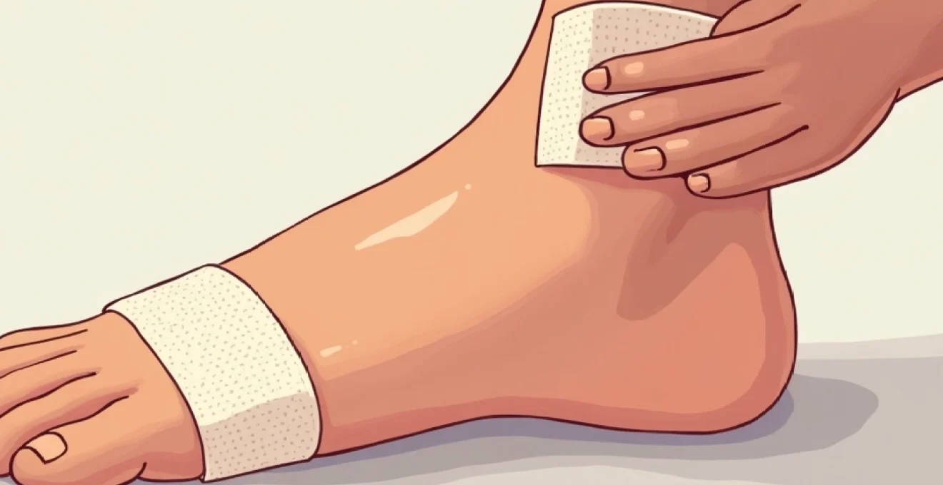
Latex adhesive contact dermatitis presents with a distinctive clinical pattern that typically follows the exact outline of the bandage or plaster application site. The inflammatory response demonstrates remarkable precision, creating sharply demarcated borders that correspond to areas of direct skin contact with latex-containing adhesives. This geometric distribution pattern serves as a crucial diagnostic clue, distinguishing latex-induced dermatitis from other inflammatory skin conditions such as eczema or irritant contact dermatitis from non-allergenic sources.
Initial clinical manifestations usually begin with erythema and mild oedema at the contact site, progressing to more pronounced inflammatory changes over subsequent hours. Patients frequently report intense pruritus that may persist for several days after bandage removal, often accompanied by a burning or stinging sensation. The temporal relationship between adhesive application and symptom onset provides valuable diagnostic information, particularly when symptoms consistently occur with latex-containing products but not with latex-free alternatives.
Erythematous papular eruptions and vesicular formations
Erythematous papular eruptions represent the most common early manifestation of latex adhesive contact dermatitis, typically appearing within 12-24 hours of initial exposure. These raised, inflammatory lesions develop as a result of dermal inflammatory cell infiltration and localised vasodilation. The papules often exhibit a characteristic red-to-purple colouration and may demonstrate central scaling or crusting as the reaction progresses. Patients frequently describe these lesions as intensely pruritic, leading to secondary excoriation and potential bacterial superinfection.
Vesicular formations occur in approximately 30-40% of latex contact dermatitis cases and represent a more severe inflammatory response. These fluid-filled lesions develop when inflammatory mediators cause significant capillary permeability and dermal oedema. Vesicles typically measure 2-5mm in diameter and contain clear to slightly turbid fluid rich in inflammatory cells and protein. Rupture of vesicles can lead to weeping, crusting, and increased risk of secondary infection, particularly in immunocompromised patients or those with compromised skin barrier function.
Pruritic eczematous lesions with scaling patterns
Pruritic eczematous lesions develop in cases of repeated or prolonged latex exposure, representing a chronic inflammatory response characterised by epidermal hyperplasia and dermal inflammatory cell infiltration. These lesions exhibit the classic features of chronic contact dermatitis, including lichenification, scaling, and fissuring. The eczematous changes typically follow a pattern of acute inflammatory response transitioning to subacute and chronic phases, with each stage demonstrating distinct histological and clinical characteristics.
Scaling patterns in latex-induced eczematous dermatitis often demonstrate a characteristic peripheral distribution, with the most pronounced changes occurring at the borders of the original contact site. This peripheral accentuation reflects the higher concentration of latex proteins typically found at adhesive margins and the mechanical irritation caused by bandage edges. Chronic scratching due to persistent pruritus contributes to the development of secondary changes including hyperpigmentation, skin thickening, and altered barrier function that may persist long after the initial allergic response has resolved.
Chronic lichenification from prolonged latex exposure
Chronic lichenification represents the end-stage manifestation of repeated latex adhesive exposure, characterised by significant epidermal thickening, enhanced skin markings, and altered pigmentation. This condition develops through a complex interplay of chronic inflammation, mechanical trauma from scratching, and impaired barrier function. Lichenified areas typically demonstrate a leather-like texture with prominent skin markings and may exhibit hyperpigmentation or hypopigmentation depending on individual skin type and the severity of the inflammatory response.
The pathophysiology of lichenification involves chronic activation of keratinocytes and fibroblasts, leading to increased collagen deposition and epidermal hyperproliferation. Patients with chronic lichenification often experience persistent symptoms including pruritus, burning sensations, and skin tightness that can significantly impact quality of life. Treatment of lichenified lesions requires a comprehensive approach including potent topical corticosteroids, barrier repair agents, and strict latex avoidance to prevent further progression of skin changes.
Systemic allergic contact dermatitis spread beyond application site
Systemic allergic contact dermatitis represents a concerning complication of latex adhesive allergy where inflammatory changes extend beyond the original contact site to involve distant skin areas. This phenomenon, also known as id reaction or autosensitisation, occurs in approximately 10-15% of severe latex contact dermatitis cases and suggests significant systemic immune activation. The mechanism involves haematogenous or lymphatic spread of inflammatory mediators and possibly latex allergens, resulting in widespread eczematous eruptions that may involve the hands, face, and other body regions.
Clinical manifestations of systemic spread include symmetric eczematous lesions on the hands and forearms, facial oedema and erythema, and generalised pruritus. These systemic reactions typically develop 3-7 days after the initial contact dermatitis and may persist for several weeks despite removal of the offending latex product. Recognition of systemic spread is crucial as it indicates severe sensitisation and increased risk of future severe reactions, necessitating comprehensive latex avoidance strategies and possible immunosuppressive therapy.
Cross-reactivity patterns with hevea brasiliensis allergens
Cross-reactivity patterns with Hevea brasiliensis allergens represent a complex immunological phenomenon that extends the clinical significance of latex allergy beyond direct contact with rubber products. The natural rubber latex contains over 250 different proteins, with approximately 13 major allergens designated as Hev b 1 through Hev b 13. These allergens share structural similarities with proteins found in various plant species, leading to cross-reactive allergic responses in sensitised individuals. Understanding these cross-reactivity patterns is essential for comprehensive patient management and prevention of unexpected allergic reactions.
The prevalence of latex-associated cross-reactivity varies significantly among different populations, with studies indicating that 30-50% of latex-allergic individuals demonstrate concurrent food allergies. Healthcare workers with occupational latex exposure show particularly high rates of cross-reactivity, with some studies reporting up to 70% prevalence of associated food allergies. Environmental factors including geographic location, dietary habits, and genetic predisposition influence the development and severity of cross-reactive responses, making individualised assessment crucial for effective management strategies.
Hev b 1-13 protein Cross-Reactions with food allergens
The 13 major latex allergens demonstrate varying degrees of cross-reactivity with food proteins, with Hev b 6.02 (prohevein) and Hev b 7 (patatin-like protein) showing the strongest associations with food allergies. Hev b 6.02 shares structural homology with chitinases found in bananas, avocados, and chestnuts, while Hev b 7 demonstrates cross-reactivity with patatin proteins in potatoes and other solanaceous plants. These molecular similarities explain the high frequency of food allergies observed in latex-sensitised individuals and the potential for severe systemic reactions following consumption of cross-reactive foods.
Research has identified specific IgE binding patterns that help predict which latex-allergic patients are most likely to develop food allergies. Patients with high levels of Hev b 6.02-specific IgE demonstrate the strongest associations with tropical fruit allergies, while those with predominant Hev b 7 sensitisation show increased reactivity to potatoes and tomatoes. Component-resolved diagnostics using purified latex allergens provides valuable information for risk stratification and dietary counselling, allowing for more personalised management approaches that balance nutritional needs with allergy prevention.
Banana-avocado-kiwi-latex syndrome manifestations
Banana-avocado-kiwi-latex syndrome represents the most well-documented cross-reactive food allergy complex, affecting approximately 35-45% of latex-allergic individuals. The syndrome results from shared allergenic proteins, particularly chitinases and class I chitinase-related proteins, that demonstrate remarkable structural conservation across these plant species. Clinical manifestations range from mild oral allergy syndrome with localised mouth and throat symptoms to severe systemic anaphylactic reactions requiring emergency intervention.
Diagnostic evaluation of suspected banana-avocado-kiwi-latex syndrome requires comprehensive assessment including detailed dietary history, specific IgE testing for individual food allergens, and component-resolved diagnostics when available. Patients typically develop food allergies secondary to latex sensitisation, though the reverse pattern can occasionally occur. Management strategies involve strict avoidance of known trigger foods, emergency action plan development, and regular monitoring for expansion of cross-reactive responses to include additional fruits or vegetables within the same allergenic families.
Spina bifida patients and High-Risk latex sensitisation
Spina bifida patients represent the highest-risk population for latex sensitisation, with prevalence rates reaching 60-70% in some studies. This extraordinary susceptibility results from early and repeated exposure to latex-containing medical devices during multiple surgical procedures, bladder catheterisations, and routine medical care throughout infancy and childhood. The combination of immature immune system development and extensive mucosal and surgical site exposure creates optimal conditions for latex protein sensitisation and subsequent allergic responses.
The clinical consequences of latex allergy in spina bifida patients can be catastrophic, with documented cases of severe intraoperative anaphylaxis resulting in cardiac arrest and death. Preventive strategies implemented in paediatric neurosurgery and urology programs have significantly reduced the incidence of new latex sensitisation cases. These protocols include exclusive use of latex-free medical products, environmental latex elimination in patient care areas, and comprehensive staff education regarding latex allergy recognition and management.
Healthcare workers occupational sensitisation patterns
Healthcare workers demonstrate unique occupational sensitisation patterns that reflect both the intensity and route of latex exposure encountered in clinical practice. Studies indicate that surgical personnel, intensive care staff, and laboratory workers show the highest sensitisation rates, correlating with both direct skin contact and inhalational exposure to latex proteins. The introduction of powder-free latex gloves and subsequent transition to non-latex alternatives has dramatically reduced new sensitisation cases, though existing allergic healthcare workers continue to face occupational challenges.
Occupational sensitisation typically follows a predictable pattern beginning with irritant contact dermatitis from frequent glove use, progressing to allergic contact dermatitis, and potentially culminating in immediate-type hypersensitivity reactions. Early recognition of latex sensitisation symptoms allows for intervention before progression to severe systemic reactions. Workplace accommodation strategies include assignment to latex-free work environments, provision of alternative personal protective equipment, and modification of job responsibilities to minimise exposure risk while maintaining career viability.
Patch testing protocols using TRUE test and finn chamber methods
Patch testing remains the gold standard diagnostic method for confirming allergic contact dermatitis to latex and adhesive components, providing definitive evidence of delayed-type hypersensitivity reactions. The TRUE Test (Thin-layer Rapid Use Epicutaneous test) offers a standardised, FDA-approved approach for detecting contact allergies to common allergens including rubber accelerators and preservatives found in adhesive formulations. This ready-to-use system eliminates variability in allergen preparation and concentration, ensuring consistent and reliable results across different testing facilities and practitioners.
Finn Chamber methodology represents the traditional approach to patch testing, utilising individual chambers filled with specific allergens at predetermined concentrations. This system offers greater flexibility in allergen selection and allows for testing of patient-supplied materials such as specific bandage adhesives or custom formulations. Proper patch testing technique requires careful attention to allergen preparation, application site selection, and reading schedules to ensure accurate interpretation and clinical relevance of positive reactions.
Contemporary patch testing protocols for latex allergy investigation typically include testing for natural rubber latex using latex glove extract at 0.7 mg/mL concentration, along with rubber accelerators such as mercaptobenzothiazole, thiuram mix, and carba mix. Additional testing may include adhesive components like colophonium, p-tert-butylphenol formaldehyde resin, and various preservatives depending on the specific products suspected of causing reactions. Test interpretation requires expertise in distinguishing true allergic responses from irritant reactions, with positive results demonstrating clinical relevance through correlation with patient history and exposure patterns.
Latex-free adhesive alternatives and hypoallergenic formulations
The development of latex-free adhesive alternatives has revolutionised the management of patients with latex allergies, providing effective wound care options without the risk of allergic reactions. Modern adhesive technologies utilise synthetic polymers and advanced chemical formulations that maintain strong adhesion properties while eliminating natural rubber latex proteins. These innovations have enabled healthcare providers to deliver comprehensive care to latex-allergic patients without compromising treatment efficacy or patient safety.
Market analysis indicates that latex-free medical adhesives now comprise over 80% of the global medical adhesive market, reflecting both increased awareness of latex allergy risks and improved product performance. Regulatory requirements in many countries mandate clear labelling of latex content in medical products, facilitating safer product selection for at-risk patients. The cost differential between latex-containing and latex-free alternatives has narrowed considerably, making universal adoption of latex-free products economically feasible for most healthcare institutions.
Synthetic Polymer-Based adhesives without natural rubber
Synthetic polymer-based adhesives utilise advanced chemical technologies including polyacrylate, polyurethane, and polyisobutylene formulations that provide excellent adhesion without natural rubber latex proteins. These synthetic systems demonstrate superior biocompatibility profiles with reduced risk of both allergic contact dermatitis and skin irritation. Polyacrylate adhesives offer excellent moisture resistance and conformability, making them ideal for applications requiring extended wear times or exposure to body fluids.
Polyurethane-based adhesive systems provide exceptional flexibility and stretch recovery, accommodating joint movement and dynamic
skin applications without compromising adhesive performance. These synthetic formulations undergo extensive biocompatibility testing to ensure minimal risk of sensitisation and excellent tolerance across diverse patient populations.
Performance characteristics of synthetic polymer adhesives often exceed those of traditional latex-containing products, particularly in challenging clinical environments. Advanced formulation chemistry allows for customisation of tack, peel strength, and shear resistance to match specific clinical requirements. Quality control protocols for synthetic adhesives include rigorous testing for residual monomers, catalyst residues, and other potential sensitising agents, ensuring consistent product safety and performance.
Silicone-based medical adhesives for sensitive skin
Silicone-based medical adhesives represent the gold standard for patients with sensitive skin conditions, offering exceptional biocompatibility with minimal risk of allergic reactions or skin irritation. These adhesive systems utilise medical-grade silicone polymers that create gentle, repositionable adhesion while maintaining excellent conformability to complex anatomical contours. The unique properties of silicone adhesives include breathability, transparency, and resistance to moisture and bacterial growth, making them ideal for extended wear applications.
Clinical studies demonstrate that silicone-based adhesives reduce the incidence of medical adhesive-related skin injuries by up to 75% compared to traditional acrylic formulations. The gentle removal characteristics of silicone adhesives minimise trauma to fragile skin, particularly important in elderly patients, paediatric populations, and individuals with compromised skin integrity. Repositionability features allow healthcare providers to adjust bandage placement without losing adhesive effectiveness, improving both patient comfort and clinical outcomes.
Advanced silicone adhesive technologies incorporate antimicrobial agents and skin conditioning additives that further enhance biocompatibility and reduce infection risk. These formulations demonstrate excellent compatibility with various wound care products including hydrogels, foams, and antimicrobial dressings. Manufacturing processes for medical-grade silicone adhesives adhere to strict regulatory standards ensuring lot-to-lot consistency and freedom from potentially harmful extractable substances that could cause delayed hypersensitivity reactions.
Hydrocolloid and hydrogel dressing technologies
Hydrocolloid and hydrogel dressing technologies provide innovative adhesive-free alternatives for wound management in latex-allergic patients, utilising moisture-responsive polymers that create secure fixation through gel formation and mechanical adhesion. Hydrocolloid dressings consist of gel-forming agents such as carboxymethylcellulose, gelatin, and elastomers suspended in an adhesive matrix that becomes increasingly adherent as it absorbs wound exudate. This mechanism eliminates the need for traditional chemical adhesives while providing excellent seal formation and bacterial barrier properties.
Hydrogel dressing systems offer superior moisture management capabilities through their high water content and three-dimensional polymer networks that facilitate controlled fluid exchange. These dressings demonstrate excellent biocompatibility profiles with minimal risk of sensitisation reactions, making them particularly suitable for patients with multiple contact allergies. Cooling properties of hydrogel dressings provide additional therapeutic benefits including pain relief and reduced inflammatory response, enhancing patient comfort during the healing process.
Clinical applications of hydrocolloid and hydrogel technologies extend beyond simple wound coverage to include advanced therapeutic functions such as controlled drug delivery, growth factor incorporation, and antimicrobial activity. Modern formulations integrate silver nanoparticles, iodine complexes, or honey-based antimicrobial agents directly into the polymer matrix, providing sustained antimicrobial action without additional chemical adhesives. Research continues to advance these technologies with the development of smart dressings that change colour to indicate infection or healing progress, providing valuable clinical information without requiring dressing removal.
Topical corticosteroid treatment protocols and barrier restoration
Topical corticosteroid treatment protocols for latex adhesive contact dermatitis require careful consideration of lesion severity, anatomical location, and patient-specific factors to achieve optimal therapeutic outcomes while minimising adverse effects. The selection of appropriate corticosteroid potency depends on the acute versus chronic nature of the inflammatory response, with acute vesicular reactions typically requiring medium to high-potency preparations, while chronic lichenified lesions may benefit from ultra-high potency formulations for limited durations. Treatment protocols must balance anti-inflammatory efficacy with the risk of skin atrophy, particularly in sensitive anatomical areas such as the face, neck, and inframammary regions.
Contemporary treatment approaches emphasise combination therapy utilising topical corticosteroids alongside barrier restoration agents to address both the inflammatory component and underlying epidermal dysfunction. Sequential treatment protocols begin with appropriate-strength corticosteroids for acute inflammation control, followed by gradual transition to barrier repair moisturisers and emollient therapies to restore normal skin function. This approach reduces the risk of corticosteroid dependence while promoting long-term skin health and resilience against future allergic reactions.
Barrier restoration strategies incorporate advanced understanding of stratum corneum physiology and the role of ceramides, fatty acids, and natural moisturising factors in maintaining healthy skin barrier function. Therapeutic formulations containing physiological lipid ratios, hyaluronic acid, and peptide complexes demonstrate superior efficacy in restoring barrier integrity compared to traditional moisturising products. Patient education regarding proper application techniques, frequency of use, and recognition of treatment response ensures optimal therapeutic outcomes and prevents progression to chronic dermatitis patterns that may require more intensive interventions.
Long-term management protocols include regular dermatological follow-up to monitor treatment response and adjust therapy as needed, comprehensive latex avoidance counselling, and development of emergency action plans for patients at risk of systemic allergic reactions. Quality of life assessment forms an integral component of treatment evaluation, as chronic contact dermatitis can significantly impact daily activities, sleep quality, and psychological wellbeing. Successful management requires a multidisciplinary approach involving dermatologists, allergists, and occupational health specialists to ensure comprehensive care and prevention of recurrent allergic episodes.