The cervical spine’s C3-C4 segment represents one of the most biomechanically active regions in the neck, making it particularly susceptible to degenerative changes that can lead to disc osteophyte complex formation. This condition, characterised by the concurrent development of intervertebral disc pathology and bony overgrowths called osteophytes, affects thousands of individuals annually and can significantly impact quality of life. The C3-C4 level’s unique anatomical position and its role in supporting head movements whilst maintaining spinal stability creates an environment where degenerative processes often accelerate over time. Understanding the intricate pathophysiology, clinical presentations, and management strategies for disc osteophyte complex at this specific level is crucial for healthcare professionals and patients alike, as early recognition and appropriate intervention can prevent progression to more severe neurological complications.
Anatomical structure and pathophysiology of C3-C4 disc osteophyte complex
Cervical vertebrae C3-C4 biomechanical framework and intervertebral disc composition
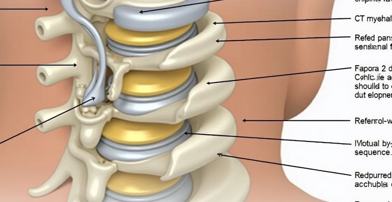
The C3-C4 spinal segment forms a critical component of the subaxial cervical spine, positioned between the highly mobile atlas-axis complex above and the more stable lower cervical vertebrae below. This intermediate location subjects the C3-C4 level to unique biomechanical stresses during daily activities. The intervertebral disc at this level consists of an outer fibrous annulus fibrosus and an inner gel-like nucleus pulposus, which work together to absorb shock and facilitate smooth cervical motion. The annulus fibrosus contains multiple concentric layers of collagen fibres arranged in alternating orientations, providing tensile strength whilst allowing controlled movement between vertebrae.
The C3-C4 disc experiences significant loading during flexion, extension, and rotational movements of the cervical spine. Unlike lumbar discs, cervical discs are relatively thin and experience higher stress concentrations per unit area. The posterior aspect of the disc is reinforced by the posterior longitudinal ligament, whilst the anterior portion is supported by the anterior longitudinal ligament. These anatomical features create a complex mechanical environment where degenerative changes often begin in the posterior annulus, particularly in areas where the annular fibres are naturally thinner and more susceptible to microscopic tears.
Osteophyte formation mechanisms in uncovertebral and facet joints
Osteophyte development at the C3-C4 level occurs through a complex cascade of inflammatory and mechanical processes. The uncovertebral joints, also known as joints of Luschka, are unique to the cervical spine and play a pivotal role in osteophyte formation. These pseudoarthroses develop between the uncinate processes of the lower vertebra and the bevelled edges of the vertebral body above. When disc degeneration begins, altered biomechanics increase stress on these joints, triggering osteoblastic activity that results in bony spur formation.
The facet joints at C3-C4 also contribute significantly to osteophyte complex development. These synovial joints, oriented at approximately 45 degrees to the horizontal plane, experience increased loading when disc height decreases. The resulting joint space narrowing leads to cartilage degeneration, subchondral sclerosis, and eventual osteophyte formation along the joint margins. This process is mediated by inflammatory cytokines, including interleukin-1 and tumour necrosis factor-alpha, which stimulate bone and cartilage remodelling pathways.
Degenerative cascade leading to Disc-Osteophyte complex development
The pathogenesis of disc osteophyte complex at C3-C4 follows a predictable degenerative cascade that typically begins in the fourth or fifth decade of life. Initial changes occur within the nucleus pulposus, where proteoglycan content decreases and water retention capacity diminishes. This biochemical alteration leads to reduced disc height and increased stress on the annulus fibrosus. Microscopic tears develop in the posterior annulus, creating pathways for nucleus material to migrate toward the spinal canal or neural foramina.
As disc degeneration progresses, the body’s natural repair mechanisms become activated. Fibroblasts proliferate within the damaged annular tissue, laying down disorganised collagen in an attempt to restore structural integrity. Simultaneously, the decreased disc height alters load distribution across the vertebral endplates, leading to reactive bone formation. This osteoblastic response results in endplate sclerosis and the formation of osteophytes along the vertebral margins. The combination of disc material displacement and osteophyte growth creates the characteristic appearance of disc osteophyte complex on imaging studies.
Neural foramen stenosis and spinal canal narrowing pathways
The development of neural foramen stenosis at C3-C4 represents a critical pathway through which disc osteophyte complex becomes clinically significant. The neural foramen, measuring approximately 4-6 millimetres in diameter at this level, provides passage for the C4 nerve root and accompanying vessels. Osteophyte formation from the uncovertebral joints posterolaterally and facet joint hypertrophy posteriorly create a pincer-like compression mechanism that gradually narrows the foramen.
Disc osteophyte complex at C3-C4 often involves extension of disc material beyond the confines of the vertebral margins, accompanied by osteophyte formation that further compromises available space for neural structures.
Spinal canal narrowing occurs through a similar but distinct mechanism. Posterior osteophyte formation, combined with ligamentum flavum hypertrophy and potential disc herniation, reduces the effective diameter of the spinal canal. The cervical spinal canal normally measures 17-18 millimetres in anteroposterior diameter at the C3-C4 level. When this dimension decreases to less than 13 millimetres, the risk of spinal cord compression increases significantly, potentially leading to myelopathic symptoms.
Clinical manifestations and neurological symptoms of C3-C4 disc osteophyte complex
Cervical radiculopathy patterns and C4 nerve root compression syndromes
C4 radiculopathy presents with distinct clinical features that differentiate it from other cervical nerve root compression syndromes. The C4 nerve root primarily innervates the diaphragm through the phrenic nerve, though it also contributes to sensation over the lower neck and upper shoulder region. Patients with C4 nerve root compression may experience referred pain patterns that extend from the suboccipital region down to the superior aspect of the shoulder girdle. Unlike lower cervical radiculopathies, C4 compression rarely causes significant motor weakness in the upper extremities.
The sensory distribution of the C4 dermatome encompasses the lower posterior neck, extending laterally over the acromioclavicular joint and superior shoulder region. Patients often describe a deep, aching sensation that may be accompanied by burning or tingling sensations. The pain typically worsens with cervical extension and rotation toward the affected side, as these movements further narrow the neural foramen. Interestingly, some patients report respiratory symptoms such as shortness of breath or a sensation of incomplete inspiration, reflecting the C4 nerve root’s contribution to diaphragmatic innervation.
Myelopathic symptoms secondary to spinal cord compression
When disc osteophyte complex at C3-C4 causes significant spinal canal stenosis, patients may develop cervical myelopathy, a serious condition requiring urgent attention. Early myelopathic symptoms are often subtle and may be attributed to normal ageing processes. Patients typically report difficulty with fine motor tasks such as buttoning clothing, writing, or manipulating small objects. This reflects compression of the lateral corticospinal tracts, which control fine motor function in the hands.
Progressive myelopathy manifests with gait disturbances characterised by spastic gait patterns , where patients walk with a wide-based, unsteady gait and may experience frequent falls. The Lhermitte’s sign, described as an electric shock-like sensation radiating down the spine and into the extremities with neck flexion, may be positive in cases of significant cord compression. Advanced myelopathy can lead to bowel and bladder dysfunction, though this is more commonly associated with lower cervical or thoracic cord compression.
Referred pain patterns to shoulder, scapular, and upper extremity regions
The complex innervation patterns of the cervical spine create opportunities for referred pain phenomena that can make diagnosis challenging. Disc osteophyte complex at C3-C4 frequently generates pain that radiates to the ipsilateral shoulder and scapular region, often mimicking rotator cuff pathology or other shoulder disorders. This occurs through convergence of nociceptive pathways in the dorsal horn of the spinal cord, where cervical afferents share neural pathways with those from the shoulder region.
Patients may describe a deep, gnawing pain beneath the medial border of the scapula that worsens with prolonged sitting or computer work. The pain often has a positional component, improving with changes in head and neck posture. Some individuals report that the pain awakens them from sleep, particularly when sleeping in prone positions that hyperextend the cervical spine. The referred nature of this pain can lead to misdiagnosis and inappropriate treatment of peripheral structures rather than addressing the underlying spinal pathology.
Motor weakness and sensory deficits in C4 dermatome distribution
While motor weakness is less prominent in C4 radiculopathy compared to lower cervical nerve root compressions, subtle deficits may be detectable on careful examination. The most significant motor contribution of the C4 nerve root is to diaphragmatic function via the phrenic nerve. Patients with severe C4 compression may demonstrate reduced diaphragmatic excursion on pulmonary function testing or fluoroscopic evaluation. This can manifest clinically as exercise intolerance or dyspnoea on exertion, particularly during activities requiring increased respiratory effort.
Sensory deficits in the C4 distribution typically involve decreased sensation to light touch and pinprick over the lower posterior neck and superior shoulder region. These deficits may be subtle and require careful comparison between affected and unaffected sides. The sensory examination should include assessment of vibration and position sense, as these modalities may be affected in cases with mild spinal cord compression. Documentation of sensory mapping is crucial for monitoring progression and treatment response over time.
Advanced imaging techniques for C3-C4 disc osteophyte complex assessment
MRI T2-Weighted sagittal and axial sequences for disc degeneration grading
Magnetic resonance imaging represents the gold standard for evaluating disc osteophyte complex at C3-C4, with T2-weighted sequences providing optimal contrast for assessing disc morphology and hydration status. Sagittal T2-weighted images allow for comprehensive evaluation of disc height, signal intensity, and the presence of posterior disc bulging or herniation. Normal cervical discs demonstrate bright, homogeneous signal intensity on T2-weighted sequences, reflecting high water content within the nucleus pulposus.
Degenerative changes appear as progressive signal loss on T2-weighted images, often beginning centrally within the nucleus and extending peripherally as degeneration advances. The Pfirrmann grading system, originally developed for lumbar discs but applicable to cervical pathology, provides a standardised method for quantifying degenerative changes. Grade 1 represents normal disc morphology with bright, homogeneous signal, whilst Grade 5 indicates severe degeneration with dark signal intensity and collapsed disc space. Axial T2-weighted images complement sagittal sequences by providing detailed visualisation of foraminal narrowing and the relationship between disc material and neural structures.
CT myelography protocol for detailed osteophyte morphology analysis
Computed tomography myelography offers superior visualisation of bony anatomy and osteophyte morphology compared to conventional MRI, making it particularly valuable for surgical planning in complex cases. The procedure involves intrathecal injection of contrast material followed by high-resolution CT imaging, providing excellent delineation of the spinal cord, nerve roots, and surrounding bony structures. This technique is especially useful when MRI is contraindicated or when detailed assessment of bony pathology is required.
The myelographic contrast outlines the subarachnoid space, clearly demonstrating areas of neural compression by disc osteophyte complexes. Posterior osteophytes appear as bony excrescences that indent the contrast column, whilst disc material may create soft tissue impressions on the thecal sac. Three-dimensional reconstructions can provide surgeons with detailed anatomical roadmaps, facilitating precise preoperative planning and reducing intraoperative complications. The dynamic component of CT myelography, performed in flexion and extension, can reveal instability patterns that may influence treatment decisions.
Dynamic Flexion-Extension radiographs for cervical instability evaluation
Dynamic radiographic evaluation plays a crucial role in assessing cervical spine stability at the C3-C4 level, particularly in patients with advanced degenerative changes. Standard lateral flexion and extension radiographs can demonstrate abnormal motion patterns that may not be apparent on static imaging studies. The normal range of flexion-extension motion at C3-C4 is approximately 15-20 degrees, with translation of less than 2 millimetres considered physiological.
Degenerative instability may manifest as excessive angular motion, abnormal translation patterns, or paradoxical motion during flexion-extension manoeuvres. The presence of retrolisthesis or anterolisthesis exceeding 2 millimetres suggests ligamentous insufficiency and may influence surgical decision-making. Dynamic imaging is particularly important in patients being considered for motion-preserving procedures, as the presence of instability may contraindicate such interventions in favour of fusion techniques.
Quantitative MRI biomarkers using pfirrmann and modic classification systems
Advanced MRI analysis incorporates standardised classification systems that provide objective measures of degenerative changes and help guide treatment decisions. The Pfirrmann classification system grades disc degeneration based on signal intensity, disc height, and morphological characteristics visible on T2-weighted sagittal images. This system demonstrates excellent inter-observer reliability and correlates well with histological findings of disc degeneration.
Modic changes represent vertebral endplate abnormalities that often accompany disc degeneration and may influence clinical symptoms and treatment outcomes in patients with disc osteophyte complex.
Modic changes appear as signal alterations in the vertebral bone marrow adjacent to degenerating discs. Type 1 changes, characterised by low signal on T1-weighted and high signal on T2-weighted images, represent bone marrow oedema and inflammation. Type 2 changes show high signal on both T1 and T2-weighted sequences, indicating fatty infiltration of the bone marrow. Type 3 changes, appearing as low signal on both sequences, represent subchondral sclerosis. The presence and type of Modic changes can provide insights into the inflammatory status of the disc osteophyte complex and may influence treatment strategies.
Conservative treatment protocols and interventional pain management
Conservative management represents the initial treatment approach for most patients with disc osteophyte complex at C3-C4, particularly those without severe neurological deficits or progressive myelopathy. The foundation of conservative care involves activity modification, with patients advised to avoid positions and movements that exacerbate symptoms. Cervical extension and prolonged static postures, such as computer work or reading, often worsen symptoms by further narrowing the neural foramina. Ergonomic modifications to workstations and sleeping arrangements can provide significant symptom relief.
Pharmacological interventions form a crucial component of conservative management. Non-steroidal anti-inflammatory drugs (NSAIDs) address both pain and inflammation associated with disc osteophyte complex. Ibuprofen, naproxen, or diclofenac are commonly prescribed, with dosing adjusted based on patient tolerance and renal function. For patients with contraindications to NSAIDs, acetaminophen provides analgesic benefits without anti-inflammatory effects. Muscle relaxants such as cyclobenzaprine or tizanidine may be beneficial for patients experiencing cervical muscle spasm secondary to their underlying pathology.
Physical therapy interventions target multiple aspects of disc osteophyte complex pathophysiology. Manual therapy techniques, including cervical mobilisation and manipulation, can improve segmental mobility and reduce muscle
spasm through improved cervical mechanics. Cervical traction, whether performed manually or with mechanical devices, can temporarily increase foraminal dimensions and decompress neural structures. The effectiveness of traction varies among patients, with some experiencing significant symptom relief whilst others derive minimal benefit.
Postural correction exercises targeting the deep cervical flexors and upper thoracic extensors help address the forward head posture commonly associated with cervical spine pathology. Strengthening exercises for the cervical stabilising muscles, including the longus colli and longus capitis, can improve dynamic stability and reduce stress on the C3-C4 segment. Patients are typically instructed in a home exercise program emphasising gentle range-of-motion exercises and isometric strengthening techniques that can be performed throughout the day.
Interventional pain management procedures offer targeted relief for patients who fail to respond adequately to conservative measures. Cervical epidural steroid injections delivered via the interlaminar or transforaminal approach can provide significant pain reduction by addressing inflammation around compressed neural structures. The procedure involves fluoroscopically guided injection of corticosteroids and local anaesthetic into the epidural space, directly targeting the pathological process. Studies demonstrate that approximately 60-70% of patients experience meaningful pain relief lasting 3-6 months following epidural injection.
Selective nerve root blocks at the C4 level can serve both diagnostic and therapeutic purposes. These procedures involve precise injection of medication around the exiting nerve root, confirming the source of symptoms whilst providing therapeutic benefit. The technique requires careful fluoroscopic guidance to avoid injury to the vertebral artery, which courses through the transverse foramina at this level. For patients with persistent symptoms despite epidural interventions, radiofrequency ablation of the medial branch nerves innervating the C3-C4 facet joints may provide longer-lasting relief by interrupting pain transmission pathways.
Surgical intervention strategies for refractory C3-C4 disc osteophyte complex
Surgical intervention becomes necessary when conservative management fails to provide adequate symptom relief or when patients develop progressive neurological deficits. The decision to proceed with surgery requires careful consideration of multiple factors, including symptom severity, functional impairment, imaging findings, and patient-specific variables such as age, activity level, and comorbidities. The presence of cervical myelopathy represents a relative surgical urgency, as delayed intervention may result in irreversible neurological damage.
Anterior cervical discectomy and fusion (ACDF) remains the gold standard surgical treatment for disc osteophyte complex at C3-C4. This approach provides direct access to the pathological disc and allows for complete removal of disc material and osteophytes compressing neural structures. The procedure involves removal of the intervertebral disc, decompression of the spinal cord and nerve roots, and placement of an interbody graft to maintain disc height and promote fusion. Fusion rates exceeding 95% are routinely achieved with modern techniques and instrumentation.
The surgical technique begins with a transverse incision along a natural skin crease, typically on the right side to avoid injury to the recurrent laryngeal nerve. Careful dissection through fascial planes provides access to the anterior cervical spine whilst preserving vital structures including the oesophagus, trachea, and carotid sheath. Intraoperative imaging confirms the correct surgical level, after which the disc is systematically removed using microsurgical techniques. Posterior osteophytes are carefully excised under magnification, with particular attention to avoid injury to the dura mater or nerve roots.
Modern interbody cage technology has revolutionised ACDF outcomes by providing immediate structural support whilst promoting biological fusion. Titanium or PEEK (polyetheretherketone) cages filled with autograft, allograft, or bone morphogenetic protein create an optimal environment for bony ingrowth. The addition of anterior cervical plates further enhances fusion rates and provides immediate stability, allowing for earlier mobilisation and improved patient satisfaction. Studies demonstrate that plated constructs achieve fusion rates of 96-98% compared to 85-90% with standalone cages.
Cervical disc arthroplasty represents an alternative surgical approach that preserves motion at the treated level whilst addressing the underlying pathology. This technique involves replacement of the diseased disc with an artificial disc prosthesis designed to replicate normal cervical kinematics. Candidates for arthroplasty must meet strict criteria, including absence of facet joint arthropathy, normal cervical lordosis, and adequate bone quality. The theoretical advantages include preservation of adjacent level motion, reduced risk of adjacent segment disease, and more rapid return to function.
Long-term studies comparing cervical disc arthroplasty to ACDF demonstrate equivalent clinical outcomes with potentially reduced rates of adjacent segment degeneration in appropriately selected patients.
Posterior cervical approaches may be considered in cases with predominantly posterior pathology or when multilevel decompression is required. Posterior cervical foraminotomy provides targeted decompression of neural foramina without the need for fusion, preserving segmental motion. The procedure involves removal of the medial aspect of the facet joint and uncovertebral osteophytes through a minimally invasive approach. However, this technique is limited to cases without significant anterior pathology or spinal instability.
Laminoplasty represents another posterior option for patients with multilevel stenosis involving the C3-C4 segment. This technique involves creating a hinge on one side of the lamina whilst opening the opposite side, effectively enlarging the spinal canal without removing bone. The opened lamina is held in position with sutures, plates, or spacers, maintaining the posterior tension band whilst providing adequate decompression. Laminoplasty preserves more normal anatomy than laminectomy and fusion, though some patients experience reduced range of motion and axial neck pain postoperatively.
Prognosis and long-term outcomes following C3-C4 disc osteophyte complex management
The prognosis for patients with disc osteophyte complex at C3-C4 varies significantly based on the severity of symptoms at presentation, duration of symptoms, and chosen treatment approach. Patients with radiculopathy typically experience more favourable outcomes compared to those with myelopathy, as nerve root compression is generally more reversible than spinal cord injury. Early recognition and appropriate treatment of cervical myelopathy are crucial, as delayed intervention may result in permanent neurological deficits even after successful decompression.
Conservative management achieves satisfactory symptom relief in approximately 40-60% of patients with disc osteophyte complex at 12-month follow-up. Factors associated with successful conservative treatment include younger age, shorter symptom duration, absence of significant motor weakness, and mild-to-moderate imaging findings. Patients who respond well to initial conservative measures often maintain their improvement long-term, though periodic symptom recurrence may occur requiring additional treatment episodes. Activity modification and adherence to prescribed exercise programs play crucial roles in maintaining long-term benefits from conservative care.
Surgical outcomes following ACDF for C3-C4 disc osteophyte complex demonstrate excellent results in appropriately selected patients. Studies report good-to-excellent clinical outcomes in 85-95% of patients, with significant improvements in pain scores, functional capacity, and quality-of-life measures. Fusion rates consistently exceed 95% with modern techniques, and pseudarthrosis requiring revision surgery occurs in fewer than 5% of cases. The majority of patients return to their previous activity levels within 3-6 months postoperatively, though some restrictions on heavy lifting and high-impact activities may persist.
Adjacent segment disease represents a significant long-term concern following cervical fusion procedures, with radiographic changes developing in 15-25% of patients over 5-10 years. However, symptomatic adjacent segment disease requiring surgical intervention occurs in only 5-10% of patients, and many radiographic changes remain clinically silent. Risk factors for adjacent segment disease include advanced age, multilevel fusion, and pre-existing degenerative changes at adjacent levels. Regular follow-up imaging and clinical monitoring allow for early detection and management of adjacent segment pathology when it occurs.
Cervical disc arthroplasty outcomes demonstrate non-inferiority to fusion for appropriately selected patients, with the theoretical advantage of preserved motion potentially reducing adjacent segment disease rates. Long-term studies extending to 10 years post-implantation show maintained clinical improvement and prosthetic function in the majority of patients. However, device-related complications including wear, loosening, or heterotopic ossification can occur, and revision surgery may be technically challenging compared to fusion procedures.
Patient selection criteria and surgical technique continue to evolve as long-term outcome data becomes available for motion preservation technologies in cervical spine surgery.
Functional outcomes following treatment of C3-C4 disc osteophyte complex are generally favourable, with most patients experiencing significant improvement in pain and disability scores. The Neck Disability Index (NDI) and Visual Analogue Scale (VAS) scores typically improve by 60-80% following successful surgical intervention. Return-to-work rates exceed 85% for patients engaged in sedentary occupations, whilst those performing heavy manual labour may require job modifications or career changes. Patient-reported outcome measures consistently demonstrate high satisfaction rates with both conservative and surgical management approaches when appropriately applied.
Long-term quality of life outcomes reveal that successful treatment of disc osteophyte complex can restore near-normal function and activity levels. Patients often report improved sleep quality, increased exercise tolerance, and reduced dependence on pain medications following effective treatment. However, some individuals may experience residual symptoms or activity limitations, particularly those with advanced degenerative changes or prolonged symptom duration prior to treatment. Ongoing research continues to refine treatment algorithms and improve outcomes for this challenging condition affecting the cervical spine’s critical C3-C4 segment.