Fingernail curvature represents one of the most telling indicators of underlying health conditions, with downward curling at the tips serving as a critical diagnostic marker that healthcare professionals closely monitor. This phenomenon, ranging from subtle changes barely noticeable to the untrained eye to dramatic alterations that fundamentally reshape the nail’s appearance, can signal everything from nutritional deficiencies to serious systemic diseases. Understanding the intricate mechanisms behind nail curvature becomes essential for early detection of potential health issues, as the nail matrix responds sensitively to changes in blood chemistry, oxygen levels, and cellular metabolism. The relationship between nail morphology and systemic health has been recognised for centuries, yet modern dermatological research continues to unveil new connections between specific curvature patterns and underlying pathophysiology.
Nail matrix dysfunction and koilonychia development
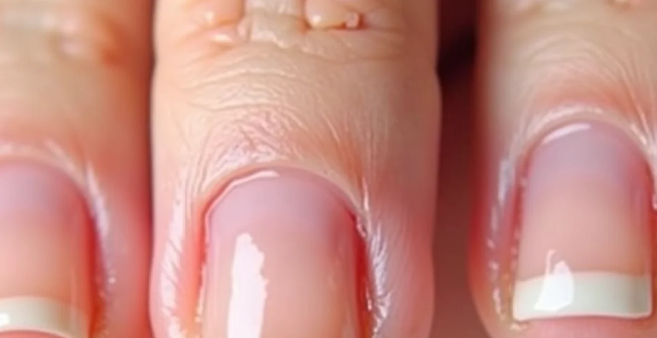
The nail matrix serves as the primary site of nail production, functioning as a highly specialised tissue that orchestrates the complex process of keratin synthesis and nail plate formation. When this delicate structure experiences dysfunction, the resulting nail abnormalities can manifest as various forms of curvature, with koilonychia representing one of the most clinically significant presentations. This condition, characterised by nails that curve upward at the edges while becoming concave in the centre, creates a distinctive spoon-like appearance that often indicates underlying systemic health issues.
Longitudinal ridging and cellular keratin production disorders
Longitudinal ridging frequently accompanies nail curvature disorders, reflecting disruptions in the normal keratinocyte differentiation process within the nail matrix. These ridges develop when the orderly progression of cells from the matrix to the nail plate becomes irregular, creating alternating bands of varying thickness that contribute to overall nail instability. The cellular keratin production process relies heavily on adequate protein synthesis, vitamin availability, and mineral cofactors, with deficiencies in any of these components leading to compromised nail structure. Keratin disorders can manifest as both horizontal and vertical ridging patterns, each indicating different underlying pathophysiological processes affecting nail matrix function.
Lunula changes and proximal nail fold abnormalities
The lunula, or the visible portion of the nail matrix appearing as a white crescent at the nail base, provides valuable insights into matrix health and function. Changes in lunula size, colour, or visibility often precede more obvious nail curvature abnormalities, serving as early warning indicators of developing matrix dysfunction. Proximal nail fold abnormalities frequently accompany these lunular changes, with inflammation, thickening, or colour alterations reflecting the broader systemic processes affecting nail growth. The proximal nail fold serves multiple protective functions while housing the stem cell populations responsible for continuous nail regeneration.
Onychocyte differentiation patterns in Spoon-Shaped nails
Onychocytes, the specialised cells that form the nail plate, undergo a carefully regulated differentiation process that determines final nail structure and integrity. In spoon-shaped nails, this differentiation pattern becomes disrupted, leading to altered cell alignment and compromised structural integrity. The normal progression from proliferative basal cells to fully keratinised nail plate components requires precise timing and coordination of multiple cellular processes. Disrupted onychocyte differentiation can result from various factors, including iron deficiency, thyroid dysfunction, and autoimmune conditions that alter the cellular microenvironment within the nail matrix.
Beau’s lines formation and growth interruption mechanisms
Beau’s lines represent horizontal indentations across the nail plate that form when nail growth temporarily stops or significantly slows due to severe systemic stress or illness. These lines provide a historical record of health events, with their position on the nail indicating when the interruption occurred based on normal nail growth rates. The formation mechanism involves temporary cessation of matrix activity, followed by resumed growth that creates a visible demarcation line. Multiple Beau’s lines can indicate repeated episodes of systemic stress, while their depth correlates with the severity and duration of the underlying condition affecting nail matrix function.
Iron deficiency anaemia and platonychia manifestations
Iron deficiency anaemia represents the most common nutritional cause of nail curvature abnormalities, with the relationship between iron status and nail health being both profound and well-documented. The condition manifests through multiple pathways, affecting not only the oxygen-carrying capacity of blood but also the fundamental cellular processes required for healthy nail growth. Platonychia , characterised by flattened nail plates that may progress to spoon-shaped deformities, serves as a classic presentation of severe iron deficiency that has persisted for extended periods.
The pathophysiology underlying iron deficiency-related nail changes involves complex interactions between oxygen delivery, cellular metabolism, and protein synthesis within the nail matrix. Iron serves as a crucial cofactor for numerous enzymatic processes involved in collagen production, keratin synthesis, and cellular respiration. When iron stores become depleted, these processes become compromised, leading to the production of structurally deficient nail plates that lack the normal convex curvature and strength characteristics of healthy nails.
Ferritin level correlation with nail plate thickness
Ferritin levels provide the most accurate assessment of total body iron stores, with research demonstrating a direct correlation between serum ferritin concentrations and nail plate thickness measurements. Studies indicate that ferritin levels below 30 ng/mL consistently correlate with measurable nail thinning, while levels below 15 ng/mL typically result in visible nail curvature abnormalities. The relationship between ferritin and nail thickness follows a predictable pattern, with nail plate measurements decreasing proportionally as iron stores become more severely depleted. Nail thickness measurements can serve as a non-invasive indicator of iron status, particularly in populations where laboratory testing may not be readily available.
Haemoglobin concentration effects on nail bed vascularity
Haemoglobin concentration directly influences nail bed vascularity, with decreased oxygen-carrying capacity leading to compensatory changes in blood flow patterns that affect nail growth and structure. The nail bed contains an extensive vascular network that supplies the matrix with essential nutrients and oxygen required for optimal nail production. When haemoglobin levels drop below normal ranges, this vascular network must work harder to maintain adequate tissue oxygenation, often resulting in visible changes to nail bed colour and nail growth patterns. Chronic anaemia can lead to permanent alterations in nail bed architecture, contributing to persistent nail curvature abnormalities even after iron levels have been restored.
Transferrin saturation and keratinocyte health
Transferrin saturation reflects the body’s ability to transport iron effectively to tissues, with levels below 20% indicating functional iron deficiency that can significantly impact keratinocyte health within the nail matrix. Keratinocytes require adequate iron availability for optimal protein synthesis and cellular differentiation processes that determine final nail structure. Transferrin saturation measurements often provide earlier indication of developing iron deficiency than haemoglobin levels alone, making this parameter particularly valuable for identifying nail-related iron deficiency in its early stages. The relationship between transferrin saturation and nail health demonstrates the importance of not just total iron stores but also iron bioavailability for maintaining normal nail structure.
Plummer-vinson syndrome associated nail dystrophy
Plummer-Vinson syndrome represents a severe manifestation of chronic iron deficiency that produces characteristic nail dystrophy alongside other systemic symptoms including dysphagia and oral mucosal changes. The nail changes associated with this syndrome typically include severe koilonychia, increased nail fragility, and unusual growth patterns that may persist even after iron replacement therapy. This condition demonstrates the long-term consequences of untreated iron deficiency on nail matrix function, with some changes becoming irreversible due to permanent structural alterations in the nail-producing tissues. The nail dystrophy in Plummer-Vinson syndrome serves as an important diagnostic marker that can help identify this potentially serious condition before more severe complications develop.
Occupational chemical exposure and nail deformation
Occupational chemical exposure represents an increasingly recognised cause of nail deformation, with certain industries presenting particular risks for developing characteristic nail curvature patterns. The mechanism of chemical-induced nail changes involves direct toxic effects on the nail matrix, disruption of normal keratinisation processes, and inflammatory responses that alter nail growth patterns. Understanding these occupational relationships becomes crucial for both prevention and early intervention in affected workers.
Chemical exposure can affect nails through multiple pathways, including direct contact with the nail plate and surrounding tissues, systemic absorption leading to matrix dysfunction, and inflammatory responses that alter local blood flow and nutrient delivery. The timing of exposure relative to nail growth cycles determines the specific pattern of abnormalities observed, with acute exposures typically producing discrete changes while chronic exposure leads to progressive deterioration of nail structure. Occupational nail disorders often present with characteristic patterns that can help identify the specific causative agents involved.
Petroleum-based solvent contact dermatitis
Petroleum-based solvents commonly used in industrial settings can cause significant nail damage through direct dehydration of the nail plate and surrounding tissues. These chemicals strip away natural oils and moisture that maintain nail flexibility, leading to brittleness, cracking, and eventual curvature abnormalities as the nail structure becomes compromised. Repeated exposure to petroleum solvents can result in permanent changes to nail texture and growth patterns, with some workers developing characteristic ridging and curvature that persists long after exposure cessation. The severity of damage correlates with both the concentration of solvents encountered and the duration of exposure, making proper protective equipment essential for prevention.
Alkali solution ph imbalance on nail structure
Alkali solutions with high pH levels can cause severe disruption of nail protein structure through denaturation of keratin and alteration of disulphide bonds that maintain nail integrity. Exposure to strong alkalis often results in immediate softening of the nail plate, followed by progressive weakening and eventual curvature as the structural framework becomes compromised. pH-related nail damage typically produces characteristic patterns of deterioration that begin at the exposed edges and progress toward the nail base, creating a distinctive appearance that can help identify alkali exposure as the causative factor. Recovery from alkali-induced nail damage often requires months of nail regrowth and may result in permanent changes to nail thickness and curvature patterns.
Heavy metal poisoning fingernail indicators
Heavy metal poisoning can produce distinctive nail changes that serve as important diagnostic indicators of toxic exposure, with different metals producing characteristic patterns of nail involvement. Lead poisoning typically results in transverse white lines known as Mees’ lines, while arsenic exposure can cause both horizontal banding and longitudinal ridging accompanied by nail curvature abnormalities. Mercury exposure often leads to a distinctive blue-black discolouration along with progressive nail weakening and curvature changes. The ability of nails to concentrate and retain heavy metals makes them valuable specimens for toxicological analysis, with nail clippings providing information about exposure patterns over several months.
Formaldehyde exposure and nail hardening disorders
Formaldehyde exposure, common in healthcare, laboratory, and manufacturing environments, can paradoxically cause both excessive nail hardening and eventual structural failure leading to curvature abnormalities. Initial exposure typically results in increased nail hardness and brittleness, but chronic exposure often leads to matrix damage that produces structurally deficient nail plates prone to breaking and curling. Formaldehyde-induced nail changes often present with a characteristic pattern of excessive hardness followed by sudden failure and fragmentation, creating irregular curvature patterns that differ from those seen with other chemical exposures. The long-term effects of formaldehyde exposure on nail matrix function can result in permanent alterations to nail growth patterns and structural integrity.
Autoimmune conditions affecting nail curvature
Autoimmune conditions represent a significant category of diseases that can profoundly affect nail structure and curvature through multiple pathophysiological mechanisms. These conditions typically involve immune system attacks on various tissues, including those involved in nail production and maintenance, leading to characteristic patterns of nail abnormalities that can serve as important diagnostic markers. Autoimmune nail manifestations often precede other systemic symptoms, making nail examination a valuable tool for early disease detection.
The relationship between autoimmune disease and nail curvature involves complex interactions between inflammatory mediators, altered blood flow patterns, and direct immune attacks on nail matrix tissues. Different autoimmune conditions tend to produce characteristic nail changes, with some affecting primarily the nail plate structure while others impact the supporting tissues and vascular supply. Understanding these patterns becomes crucial for healthcare providers seeking to identify underlying autoimmune disease in patients presenting with unexplained nail abnormalities.
Systemic lupus erythematosus frequently produces nail changes including curvature abnormalities, ridging, and colour alterations that reflect the broader systemic inflammatory process. Psoriasis can cause severe nail involvement with pitting, ridging, and curvature changes that may precede skin manifestations by months or years. Rheumatoid arthritis often results in nail fold inflammation and secondary nail deformities that contribute to overall nail structure compromise. Autoimmune nail patterns can provide valuable diagnostic information when evaluated alongside other clinical findings and laboratory results.
The nail changes seen in autoimmune disease often reflect the broader systemic inflammatory burden and can serve as a window into disease activity and treatment response patterns.
Thyroid dysfunction and nail growth abnormalities
Thyroid dysfunction represents one of the most significant endocrine causes of nail curvature abnormalities, with both hyperthyroidism and hypothyroidism producing characteristic patterns of nail changes that reflect the underlying metabolic disturbances. The thyroid gland’s crucial role in regulating cellular metabolism means that thyroid dysfunction affects virtually every aspect of nail growth, from matrix cell proliferation to final nail plate structure. Understanding these relationships becomes essential for recognising thyroid disease through nail examination and monitoring treatment response through nail improvement patterns.
Hypothyroidism typically produces nails that grow slowly, become thick and brittle, and may develop pronounced curvature as the metabolic processes required for normal nail formation become compromised. The reduced metabolic rate associated with hypothyroidism affects protein synthesis, cellular turnover, and blood flow to the extremities, all of which contribute to altered nail growth patterns. Thyroid-related nail changes often develop gradually over months to years, reflecting the insidious nature of thyroid dysfunction itself.
Hyperthyroidism can cause rapid nail growth accompanied by increased fragility and tendency toward curvature abnormalities as the accelerated metabolic processes outpace the body’s ability to maintain structural integrity. The increased metabolic demand associated with hyperthyroidism often leads to nutritional deficiencies that further compromise nail health, creating a complex pattern of abnormalities that may include both growth acceleration and structural defects. Thyroid eye disease, when present, can be accompanied by characteristic nail changes that help confirm the diagnosis and monitor disease progression.
The mechanism of thyroid-induced nail changes involves multiple pathways, including altered protein synthesis rates, changed blood flow patterns, and modified cellular differentiation processes within the nail matrix. Thyroid hormones directly influence keratinocyte function and proliferation, with both excess and deficiency leading to abnormal nail production. Recovery of normal nail appearance following thyroid treatment typically requires 6-12 months of normal nail growth, providing a useful marker for monitoring treatment effectiveness and metabolic normalisation.
Diagnostic dermatoscopy techniques for curved nail assessment
Dermatoscopy has revolutionised the assessment of nail curvature abnormalities, providing enhanced visualisation capabilities that allow for detailed examination of nail structure, vascular patterns, and surface characteristics that may not be apparent to naked eye examination. This non-invasive diagnostic technique enables healthcare providers to identify subtle changes in nail architecture that can provide crucial diagnostic information about underlying systemic conditions. Dermatoscopic nail examination has become an essential skill for dermatologists and other healthcare providers involved in nail disorder diagnosis and management.
The dermatoscopic examination of curved nails involves systematic evaluation of multiple structural components, including the nail plate surface, nail bed vasculature, periungual tissues, and matrix area when visible. Specific dermatoscopic features can help distinguish between different causes of nail curvature, with vascular patterns being particularly useful for identifying inflammatory conditions versus nutritional deficiencies. The ability to magnify and illuminate nail structures allows for detection of early changes that might otherwise go unnoticed until more advanced disease stages.
Advanced dermatoscopic techniques include polarised and non-polarised light examination, which can reveal different aspects of nail structure and pathology. Polarised light examination enhances visualisation of surface features and structural abnormalities, while non-polarised light better demonstrates vascular patterns and colour changes within the nail bed. Polarised dermatoscopy proves particularly valuable for assessing nail plate thickness variations and surface irregularities that contribute to curvature abnormalities.
Digital dermatoscopy allows for documentation and comparison of nail changes over time, enabling healthcare providers to track disease progression or treatment response with unprecedented precision. Sequential dermatoscopic imaging can reveal subtle improvements or deterioration in nail structure that might not be apparent during individual examinations. The ability to measure nail plate thickness, curvature angles, and vascular density using dermatoscopic imaging provides objective data that enhances diagnostic accuracy and treatment monitoring. Digital documentation of nail abnormalities proves particularly valuable for monitoring chronic conditions where changes occur gradually over extended periods.
Specialised dermatoscopic scoring systems have been developed to standardise the assessment of nail curvature abnormalities, allowing for consistent evaluation across different healthcare providers and clinical settings. These scoring systems typically incorporate multiple parameters including curvature severity, surface texture, colour changes, and vascular patterns to provide comprehensive assessment scores. The standardisation of dermatoscopic nail evaluation enables better communication between healthcare providers and more accurate tracking of treatment outcomes. Research continues to refine these scoring systems to improve their diagnostic accuracy and clinical utility for specific nail conditions.
The integration of dermatoscopy with other diagnostic modalities, including laboratory testing and clinical history, creates a comprehensive approach to nail disorder evaluation that maximises diagnostic accuracy while minimising unnecessary testing. Dermatoscopic findings can guide laboratory test selection by identifying patterns suggestive of specific underlying conditions, such as iron deficiency or thyroid dysfunction. The combination of detailed visual assessment with targeted laboratory evaluation often provides definitive diagnosis more efficiently than either approach used alone. Multimodal diagnostic approaches represent the current standard of care for complex nail disorders where multiple potential causes must be considered.
Training in dermatoscopic nail examination requires understanding of normal nail architecture variations across different populations and age groups, as well as familiarity with the wide range of pathological changes that can affect nail appearance. The learning curve for dermatoscopic nail assessment involves developing pattern recognition skills that allow rapid identification of significant abnormalities while avoiding over-interpretation of normal variations. Continuing education in dermatoscopic techniques remains essential as new applications and scoring systems continue to evolve. The investment in dermatoscopic equipment and training typically provides excellent returns through improved diagnostic accuracy and reduced need for invasive diagnostic procedures.