The wrinkled appearance of the penile glans, commonly known as the tip of the penis, represents a fascinating interplay of anatomical design, physiological adaptation, and developmental biology. This distinctive textural characteristic stems from complex structural arrangements that serve multiple functional purposes, ranging from sensitivity enhancement to protection during intimate contact. Understanding why this anatomical region exhibits its characteristic appearance requires examining the intricate layers of specialised tissue, vascular networks, and cellular arrangements that contribute to its unique morphology.
The glans penis, derived from the Latin word meaning “acorn,” showcases remarkable evolutionary adaptations that optimise both reproductive function and urinary processes. Its textured surface results from sophisticated cellular organisation patterns, hormonal influences, and mechanical factors that shape its development from embryonic stages through adult maturation. These morphological features reflect millions of years of evolutionary refinement, creating an organ system that balances sensitivity with durability.
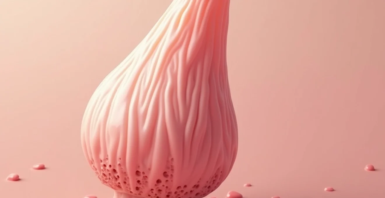
Anatomical structure of the glans penis and coronal ridge
The glans penis represents the terminal expansion of the corpus spongiosum, containing a complex network of erectile tissue, nerve endings, and specialised epithelial cells. This anatomical region exhibits distinct structural characteristics that contribute to its textured appearance, including the presence of numerous sebaceous glands, hair follicles, and vascular formations. The underlying erectile tissue creates an irregular surface topography that becomes more pronounced during different physiological states.
The coronal ridge, or corona glandis, forms the prominent circular border at the base of the glans, creating a distinctive anatomical landmark that contributes significantly to the overall textural appearance. This ridge contains concentrated nerve endings and specialised tissue arrangements that enhance sensitivity whilst creating visible surface irregularities. The junction between the glans and penile shaft exhibits varying degrees of prominence across individuals, influenced by genetic factors, hormonal exposure during development, and circumcision status.
Keratinised epithelium formation on the glans surface
The epithelial layer covering the glans undergoes varying degrees of keratinisation depending on exposure patterns and individual physiological factors. In uncircumcised males, the glans remains protected by the prepuce, maintaining a more delicate, non-keratinised epithelium that appears smoother and more sensitive. This protective environment preserves the natural mucosal characteristics of the glans surface, resulting in a softer, more pliable texture.
Conversely, circumcised males experience continuous environmental exposure that triggers adaptive keratinisation processes. This protective response creates a thicker, more resilient epithelial layer that exhibits increased surface irregularity and a more pronounced wrinkled appearance. The keratinisation process develops gradually over time, with changes becoming more noticeable during adolescence and early adulthood as hormonal influences intensify cellular turnover rates.
Coronal sulcus development and textural characteristics
The coronal sulcus, the groove separating the glans from the penile shaft, contains specialised glandular structures that contribute to surface texture variations. This anatomical depression houses numerous sebaceous glands and modified hair follicles that create microscopic surface irregularities. The accumulation of natural secretions and cellular debris within this region can temporarily enhance the wrinkled appearance, particularly in individuals with varying hygiene practices.
The depth and prominence of the coronal sulcus vary significantly among individuals, influenced by genetic factors and developmental patterns. Some men exhibit a pronounced sulcus that creates distinct visual demarcation between glans and shaft, whilst others display a more gradual transition that subtly affects overall textural perception. These variations represent normal anatomical diversity rather than pathological conditions.
Prepuce retraction effects on glans morphology
The mechanics of prepuce retraction create temporary surface texture modifications that contribute to the perceived wrinkled appearance. During retraction, the glans surface experiences mechanical stretching and compression forces that temporarily alter its topographical characteristics. Repeated retraction cycles throughout development influence the long-term textural properties of the glans epithelium.
In cases where the prepuce remains non-retractile for extended periods, the underlying glans maintains its original smooth, mucosal appearance. However, once regular retraction begins, environmental exposure initiates adaptive changes that gradually modify surface texture. This transitional period typically occurs during adolescence when hormonal changes promote increased sexual development and awareness.
Sebaceous gland distribution and fordyce spots appearance
Sebaceous glands distributed throughout the glans region contribute significantly to surface texture variations through their natural secretory activities. These microscopic structures create subtle surface elevations that become more apparent under certain lighting conditions or during states of physiological arousal. The density and activity of these glands increase during puberty under hormonal influences, potentially enhancing the wrinkled appearance.
Fordyce spots, representing enlarged sebaceous glands, commonly appear on the glans and surrounding areas as small, yellowish or white papules. These benign formations create additional surface texture variations that contribute to the overall wrinkled appearance. Their prevalence increases with age, with studies indicating that up to 85% of adult males exhibit some degree of Fordyce spot development on their genital anatomy.
Physiological factors contributing to penile glans texture
Multiple physiological mechanisms influence the textural characteristics of the penile glans, creating the distinctive wrinkled appearance that varies among individuals. Vascular dynamics play a crucial role, as the erectile tissue within the glans contains numerous blood vessels that expand and contract according to physiological demands. During flaccid states, reduced blood flow allows tissue compression that accentuates surface irregularities and creates more pronounced wrinkled patterns.
Temperature regulation also affects glans appearance, with cooler conditions promoting tissue contraction that enhances textural prominence. The glans contains specialised smooth muscle fibres that respond to temperature changes, autonomic nervous system activity, and psychological factors. These responses create dynamic surface modifications that contribute to the overall perception of wrinkled texture, particularly during non-aroused states when tissue remains in its compressed configuration.
Circumcision impact on glans keratinisation process
Circumcision fundamentally alters the glans microenvironment, triggering adaptive responses that significantly modify surface texture characteristics. The removal of the protective prepuce exposes the glans to continuous environmental contact, initiating keratinisation processes that create a thicker, more resilient epithelial layer. This adaptive response typically develops over several months to years following circumcision, with younger patients showing more rapid adaptation.
The keratinisation process involves increased production of keratin proteins within epithelial cells, creating a protective barrier that reduces sensitivity whilst enhancing durability. This cellular transformation results in visible texture changes, including increased surface roughness and more pronounced wrinkled patterns. Research indicates that circumcised males exhibit approximately 20-30% thicker epithelium compared to their uncircumcised counterparts, directly contributing to textural differences.
Smegma accumulation and surface texture alterations
Natural smegma production serves important protective and lubricating functions but can temporarily modify glans appearance when accumulation occurs. This substance consists of dead epithelial cells, sebaceous secretions, and moisture that creates a protective layer over sensitive glans tissue. Regular accumulation and removal cycles can influence long-term textural development by affecting epithelial turnover rates.
In uncircumcised males, smegma typically accumulates beneath the prepuce, creating a protective environment that maintains the glans’ natural mucosal characteristics. However, irregular hygiene practices may lead to excessive accumulation that alters surface texture perception. Proper hygiene maintenance ensures optimal glans health whilst preserving natural textural characteristics without unnecessary irritation or damage to delicate epithelial structures.
Hormonal influences on epithelial cell turnover
Testosterone and other androgens significantly influence epithelial cell turnover rates within the glans region, affecting long-term textural development. During puberty, increased hormonal activity accelerates cellular proliferation and differentiation processes that contribute to adult glans morphology. These hormonal influences continue throughout adult life, with age-related changes affecting cellular regeneration capacity and surface texture maintenance.
Dihydrotestosterone (DHT), the most potent androgen, particularly affects genital tissue development and maintenance. Higher DHT levels promote increased epithelial thickness and enhanced keratinisation, potentially creating more pronounced textural characteristics. Individual variations in hormonal sensitivity and production levels contribute to the diverse range of glans appearances observed across the male population.
Age-related changes in glans sensitivity and appearance
Advancing age brings several physiological changes that affect glans texture and overall appearance. Decreased collagen production and cellular regeneration capacity lead to subtle modifications in epithelial structure and elasticity. These age-related changes may enhance the wrinkled appearance as tissue loses some of its youthful resilience and flexibility.
Reduced hormonal production in older males affects epithelial maintenance processes, potentially leading to increased dryness and surface irregularities. However, these changes represent normal ageing processes rather than pathological conditions. Maintaining good hygiene practices and overall health can help preserve optimal glans appearance throughout the ageing process whilst accepting natural physiological modifications.
Dermatological conditions affecting glans appearance
Various dermatological conditions can significantly alter the normal textural characteristics of the glans penis, creating pathological wrinkled appearances that differ from natural anatomical variations. These conditions range from inflammatory processes to infectious diseases that modify epithelial structure and surface topography. Understanding the distinction between normal textural variations and pathological changes requires careful observation of accompanying symptoms such as pain, discolouration, or unusual discharge.
Chronic inflammatory conditions often create persistent textural modifications that may be mistaken for normal anatomical features. These pathological changes typically develop gradually over time, making early recognition challenging for both patients and healthcare providers. Professional medical evaluation becomes essential when textural changes occur alongside other concerning symptoms or when normal hygiene practices fail to maintain optimal glans health.
Balanitis xerotica obliterans and lichen sclerosus manifestations
Balanitis xerotica obliterans (BXO), also known as lichen sclerosus et atrophicus, represents a chronic inflammatory condition that creates distinctive textural changes in the glans region. This condition typically begins with white, atrophic patches that gradually expand and create a characteristic “parchment-like” appearance. The affected tissue becomes thickened and inelastic, developing a distinctly wrinkled texture that differs significantly from normal anatomical variations.
The progression of BXO involves gradual epithelial thinning combined with dermal sclerosis that creates permanent textural modifications. Advanced cases may develop contracture and stenosis that affects both appearance and function. Early recognition and appropriate treatment can help prevent severe complications, though some textural changes may persist even after successful intervention. The condition affects approximately 0.6% of adult males, with higher prevalence rates observed in certain genetic populations.
Pearly penile papules and hirsuties coronae glandis
Pearly penile papules represent normal anatomical variants that create distinctive textural patterns around the coronal ridge. These small, dome-shaped formations appear as rows of tiny bumps that encircle the glans, creating a characteristic “beaded” appearance. Despite their sometimes concerning appearance, these structures represent benign developmental variations that occur in approximately 14-48% of adult males.
The papules typically measure 1-4 millimetres in diameter and maintain consistent size and appearance throughout adult life. Their presence may enhance the perceived wrinkled texture of the glans, particularly when viewed under magnification or specific lighting conditions. No treatment is required for these normal variants, though some men seek cosmetic intervention for aesthetic reasons. Understanding their benign nature helps reduce unnecessary anxiety about normal anatomical diversity.
Candidal balanitis effects on glans surface texture
Candidal infections create temporary but significant modifications to glans texture through inflammatory processes and epithelial disruption. The infection typically begins with subtle redness and irritation that progresses to more pronounced textural changes, including scaling, erosion, and irregular surface patterns. These pathological modifications differ markedly from normal anatomical variations and usually occur alongside other symptoms such as itching, discharge, or discomfort.
The inflammatory response associated with candidal balanitis triggers increased epithelial turnover and cellular infiltration that creates temporary wrinkled patterns. Resolution of the infection typically restores normal textural characteristics, though severe or recurrent infections may lead to persistent modifications. Proper diagnosis and treatment prevent complications whilst ensuring optimal restoration of normal glans appearance and function.
Normal anatomical variations in glans morphology
The remarkable diversity observed in normal glans morphology reflects the complex interplay of genetic, developmental, and environmental factors that influence individual anatomical expression. Studies examining large populations reveal significant variations in glans size, shape, colour, and textural characteristics, all falling within the spectrum of normal human anatomy. These variations include differences in coronal ridge prominence, surface texture patterns, vascular visibility, and overall proportional relationships with surrounding penile structures.
Research indicates that normal glans morphology encompasses a wide range of textural patterns, from relatively smooth surfaces to more pronounced wrinkled appearances, with no single configuration representing the ideal or preferred state.
Genetic factors play a fundamental role in determining individual glans characteristics, with inherited traits affecting everything from epithelial thickness to vascular arrangement patterns. Family studies suggest strong heritability for certain morphological features, though environmental influences during development can modify genetic expression. Cultural and ethnic variations also contribute to morphological diversity, with different populations exhibiting distinct patterns of normal anatomical characteristics.
The relationship between glans morphology and functional capacity remains an active area of research interest. Current evidence suggests that textural variations have minimal impact on reproductive function or sexual satisfaction, with individual preference and psychological factors playing more significant roles than anatomical configuration. Understanding this relationship helps reduce anxiety about normal anatomical variations whilst promoting healthy attitudes toward genital anatomy.
Medical assessment of glans texture abnormalities
Professional medical evaluation of glans texture abnormalities requires systematic assessment techniques that distinguish between normal variations and pathological conditions. Healthcare providers utilise visual examination, palpation, and patient history to identify concerning changes that warrant further investigation or treatment. The evaluation process considers factors such as onset timing, associated symptoms, hygiene practices, and sexual health history to develop appropriate diagnostic impressions.
Dermatological examination techniques specifically adapted for genital anatomy provide optimal assessment of textural abnormalities. These approaches include careful inspection under adequate lighting, gentle palpation to assess tissue consistency, and photographic documentation when appropriate for monitoring changes over time. Advanced diagnostic techniques such as dermatoscopy or biopsy may be necessary for complex cases where clinical examination alone cannot establish definitive diagnosis.
Patient education represents a crucial component of medical assessment, helping individuals understand the distinction between normal textural variations and pathological changes requiring intervention. Healthcare providers emphasise the importance of regular self-examination and prompt medical consultation when concerning changes develop. This educational approach promotes early detection of significant conditions whilst reducing anxiety about normal anatomical characteristics.
The integration of patient-reported symptoms with objective clinical findings guides treatment decisions and follow-up planning. Successful management requires collaboration between patient and provider to ensure optimal outcomes whilst maintaining realistic expectations about achievable results. Long-term monitoring may be necessary for certain conditions, emphasising the importance of establishing strong therapeutic relationships and clear communication channels.