Stepping out of the shower only to notice an unusual amount of dead skin flaking off your body can be disconcerting, yet this phenomenon is remarkably common and typically represents your skin functioning exactly as nature intended. The warm water and steam from your shower create optimal conditions for revealing the continuous process of cellular renewal that occurs beneath the surface of your epidermis. Understanding why this happens requires delving into the intricate biological mechanisms that govern skin cell turnover, the impact of bathing conditions on your skin’s protective barrier, and recognising when excessive desquamation might signal underlying dermatological concerns.
Understanding desquamation: the natural process of skin cell shedding
Desquamation represents one of the most fundamental processes occurring within your epidermis, involving the systematic shedding of dead skin cells from the outermost layer of your skin. This process operates continuously throughout your life, serving as your body’s primary mechanism for maintaining healthy, resilient skin whilst protecting against environmental threats. The term desquamation derives from the Latin word “desquamare,” meaning to scrape off scales, which accurately describes the microscopic reality of what occurs on your skin’s surface every moment of every day.
Keratinocyte turnover cycle and stratum corneum formation
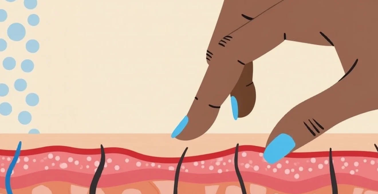
The journey of skin cell renewal begins deep within the basal layer of your epidermis, where keratinocytes undergo mitotic division to produce new cells. These newly formed keratinocytes embark on a precisely orchestrated migration upward through the various epidermal layers, gradually transforming from living, metabolically active cells into the protective, dead cells that form your stratum corneum. During this upward journey, keratinocytes synthesise increasingly large quantities of keratin proteins whilst simultaneously losing their cellular organelles and nuclei, ultimately becoming corneocytes – the flattened, protein-rich cells that provide your skin with its remarkable barrier properties.
Normal desquamation rate: 30,000-40,000 cells per minute
Your skin sheds an astounding 30,000 to 40,000 dead cells every minute, translating to approximately 40-50 million cells per day under normal physiological conditions. This remarkable rate of cellular turnover means that you completely renew your entire epidermal surface approximately every 28-30 days, though this timeframe can vary significantly based on factors such as age, health status, and environmental conditions. The majority of this cellular shedding occurs invisibly throughout the day, with individual cells detaching so gradually that they remain unnoticed. However, showering creates conditions that make this normally imperceptible process dramatically more visible, as warm water softens the connections between cells and mechanical friction accelerates their removal.
Corneodesmosomes breakdown and proteolytic enzyme activity
The orderly shedding of dead skin cells depends critically on the controlled breakdown of corneodesmosomes – specialised protein structures that maintain adhesion between adjacent corneocytes. These molecular rivets must be systematically degraded by proteolytic enzymes, particularly kallikreins and cathepsins, to allow for proper desquamation. The activity of these enzymes is carefully regulated by factors such as pH levels, humidity, and the presence of specific cofactors. When you shower, the combination of increased moisture and elevated skin temperature enhances enzymatic activity, accelerating the breakdown of corneodesmosomes and making dead cell shedding more pronounced and visible than under normal dry conditions.
Epidermal transit time: 28-day cell journey to surface
The complete journey from keratinocyte birth in the basal layer to eventual shedding from the stratum corneum typically requires 28 days in healthy adult skin, though this transit time increases with age and can be dramatically altered by various skin conditions. During the first 14 days, cells actively divide and differentiate as they move through the living layers of the epidermis. The remaining 14 days are spent in the stratum corneum, where dead cells gradually lose their intercellular attachments whilst continuing to provide protective barrier function. Understanding this timeline helps explain why changes in your health, diet, or skincare routine may not become visible for several weeks, as the cells currently on your surface began their journey nearly a month earlier.
Hot water temperature effects on epidermal barrier function
The temperature of your shower water exerts profound effects on your skin’s barrier function, with consequences that extend far beyond the immediate bathing experience. Water temperatures exceeding your skin’s optimal thermal range can trigger a cascade of biological responses that compromise barrier integrity and dramatically increase visible desquamation. Most people prefer shower temperatures between 38-42°C, yet temperatures above 40°C begin to cause measurable damage to the epidermal barrier, whilst temperatures below 37°C may not effectively remove surface debris and accumulated dead cells.
Lipid bilayer disruption through thermal damage
Excessive heat exposure disrupts the carefully organised lipid bilayers that form the mortar between your skin cells, fundamentally compromising the barrier that prevents water loss and protects against environmental irritants. These intercellular lipids, composed primarily of ceramides, cholesterol, and fatty acids, exist in a delicate crystalline arrangement that provides optimal barrier function at normal body temperature. When exposed to temperatures significantly above 40°C, these lipid structures begin to lose their organised arrangement, creating microscopic gaps that allow increased water penetration and enhanced removal of loosely attached dead cells. This thermal disruption explains why you notice substantially more dead skin after particularly hot showers compared to lukewarm bathing experiences.
Ceramide depletion and natural moisturising factor loss
Hot water accelerates the depletion of ceramides and natural moisturising factors (NMF) from your stratum corneum, creating conditions that promote excessive desquamation. Ceramides represent approximately 50% of the intercellular lipids in healthy skin and play crucial roles in maintaining barrier function and preventing excessive water loss. Simultaneously, NMF – comprising amino acids, urea, lactic acid, and other small molecules – helps maintain optimal hydration levels within the stratum corneum. High-temperature water exposure strips away these vital components more rapidly than your skin can replenish them, leading to compromised barrier function and the visible shedding of inadequately hydrated dead cells that would normally remain attached longer under optimal conditions.
Transepidermal water loss acceleration above 40°C
Scientific studies demonstrate that transepidermal water loss (TEWL) increases exponentially when skin is exposed to water temperatures exceeding 40°C, with some research showing up to 300% increases in water loss rates following hot water exposure. This accelerated water loss creates a feedback loop where compromised barrier function leads to further cellular dehydration, making dead cells more likely to detach prematurely. The increased TEWL also triggers compensatory responses within deeper epidermal layers, potentially accelerating keratinocyte turnover rates and contributing to the accumulation of dead cells ready for shedding. Understanding this relationship between temperature and water loss helps explain why post-shower skin often feels tight and appears flaky, particularly in individuals with naturally dry skin or compromised barrier function.
Sebaceous gland activity suppression in high temperature exposure
Paradoxically, whilst hot water removes existing sebum from your skin surface, it can also temporarily suppress sebaceous gland activity through thermal stress responses. This suppression reduces the natural oils that would normally help maintain stratum corneum flexibility and support proper desquamation patterns. Without adequate sebum production, dead skin cells become more brittle and prone to irregular shedding patterns, contributing to the visible flaking observed after hot showers. Recovery of normal sebaceous gland function may take several hours following thermal stress, during which time your skin remains vulnerable to excessive moisture loss and abnormal desquamation patterns.
Mechanical exfoliation during washing and towelling
The physical actions involved in washing and drying your body provide significant mechanical forces that accelerate the removal of dead skin cells beyond what would occur through natural desquamation alone. These mechanical forces work synergistically with the chemical and thermal effects of bathing to create conditions where dead cell removal becomes dramatically more apparent. Understanding the contribution of mechanical exfoliation helps explain why different washing techniques and towelling methods can substantially influence the amount of visible dead skin you observe post-shower.
Washcloths, loofahs, and even your hands create frictional forces that physically dislodge dead cells that have already lost most of their intercellular attachments but haven’t yet spontaneously shed. This mechanical action is generally beneficial, as it removes cells that might otherwise accumulate and create a dull, rough skin texture. However, excessive mechanical force can damage living cells and disrupt barrier function, leading to increased inflammation and paradoxically more visible desquamation in subsequent days.
The towelling process after showering continues this mechanical exfoliation, particularly when using traditional cotton towels with rougher textures. Vigorous rubbing with towels can remove not only loose dead cells but also cells that weren’t quite ready for natural shedding, potentially compromising barrier function. Research indicates that gentle patting motions with soft, absorbent towels minimise unnecessary mechanical trauma whilst still effectively removing excess moisture and loose cellular debris. The direction of towelling also matters, with movements that follow natural skin texture patterns causing less disruption than aggressive cross-directional rubbing.
Different body regions exhibit varying sensitivities to mechanical exfoliation, with areas like the shins, elbows, and knees typically showing more pronounced responses due to thicker stratum corneum layers and different desquamation patterns. Conversely, sensitive areas such as the face and neck require gentler mechanical forces to avoid irritation and excessive barrier disruption. Tailoring your mechanical exfoliation approach to different body regions can optimise dead cell removal whilst minimising potential adverse effects on barrier function and skin health.
Soap and cleansing agent impact on stratum corneum integrity
The cleansing agents you choose for bathing exert profound effects on stratum corneum integrity, influencing both the immediate appearance of dead skin shedding and longer-term barrier function. Modern cleansing formulations vary dramatically in their impact on skin physiology, with some products enhancing natural desquamation processes whilst others can cause significant barrier disruption and excessive cellular shedding. Understanding these effects empowers you to make informed choices about cleansing products that support optimal skin health whilst minimising unwanted side effects.
Sodium lauryl Sulphate-Induced protein denaturation
Sodium lauryl sulphate (SLS) and related surfactants commonly found in conventional soaps and body washes can cause significant protein denaturation within the stratum corneum, compromising the structural integrity of dead cells and their intercellular attachments. SLS molecules penetrate between corneocytes and disrupt the protein structures that maintain cellular cohesion, leading to premature and excessive dead cell shedding. This protein denaturation effect is dose-dependent and cumulative, meaning that regular use of high-SLS products can progressively weaken stratum corneum integrity over time. Studies have shown that SLS exposure can increase desquamation rates by up to 200% compared to gentler cleansing alternatives, explaining why individuals using conventional soaps often notice dramatically more dead skin after showering.
Ph disruption: alkaline products versus skin’s acid mantle
Your skin maintains a slightly acidic surface pH between 4.5-6.5, known as the acid mantle , which plays crucial roles in barrier function, antimicrobial defence, and proper desquamation regulation. Many traditional soaps exhibit pH levels between 8-10, creating a significant alkaline challenge that can persist for hours after cleansing. This pH disruption affects the activity of proteolytic enzymes responsible for controlled corneodesmosomes breakdown, often accelerating desquamation beyond optimal rates. Additionally, alkaline conditions can cause swelling of the stratum corneum, making dead cells more susceptible to mechanical removal and creating the appearance of increased skin shedding. pH-balanced cleansing products that respect your skin’s natural acid mantle support more controlled and appropriate desquamation patterns.
Detergent-mediated intercellular lipid extraction
Detergent molecules in cleansing products can extract vital intercellular lipids from the stratum corneum, compromising the barrier function that normally regulates desquamation timing and patterns. These lipids, including ceramides, cholesterol, and fatty acids, provide both structural support for dead cell adhesion and waterproofing functions that protect against excessive moisture penetration. When detergents remove these lipids faster than your skin can replenish them, dead cells lose their normal adhesive properties and shed more readily and visibly. This lipid extraction effect is particularly pronounced with harsh detergents and can create a cycle where barrier compromise leads to increased sensitivity and further lipid loss during subsequent cleansing sessions.
Compromised filaggrin function through chemical irritation
Filaggrin proteins play essential roles in maintaining stratum corneum structure and supporting proper desquamation patterns, yet these proteins are particularly vulnerable to chemical irritation from harsh cleansing agents. When filaggrin function becomes compromised through repeated exposure to irritating chemicals, the normal organisation of dead cells becomes disrupted, leading to irregular shedding patterns and increased visibility of desquamation. This protein degradation can also trigger inflammatory responses that further accelerate cellular turnover, creating a positive feedback loop where increased dead cell production leads to more apparent shedding. Individuals with genetic filaggrin mutations may be particularly susceptible to cleansing-related barrier disruption and excessive desquamation.
Post-shower xerosis and visible dead skin accumulation
Post-shower xerosis refers to the dry, often scaly skin condition that develops following bathing, characterised by increased visibility of dead skin cells and compromised barrier function. This condition results from the complex interplay of thermal, chemical, and mechanical factors encountered during showering, combined with the rapid evaporation of moisture from compromised skin barriers. The transition from the humid shower environment to normal ambient conditions creates additional stress on barrier function, often making dead skin accumulation more apparent in the hours following bathing.
The phenomenon of increased visible dead skin after showering represents more than simple cellular shedding – it indicates a temporary disruption in the normal desquamation process where cells that should shed gradually and invisibly instead accumulate and become apparent through clumping and irregular detachment patterns. This disruption can be influenced by numerous factors including water quality, ambient humidity, seasonal changes, and individual variations in barrier function capacity.
The visible accumulation of dead skin following showering often indicates that your skin’s natural moisture balance has been temporarily disrupted, creating conditions where normal cellular shedding becomes irregular and more apparent.
Environmental factors play significant roles in post-shower xerosis development, with low humidity conditions accelerating moisture loss and making dead cell shedding more visible. Seasonal variations affect this process, with winter months typically showing increased post-shower desquamation due to reduced ambient humidity and increased indoor heating. Air conditioning systems can create similar effects during summer months, removing moisture from both the environment and your skin surface.
Individual factors such as age, skin type, and underlying health conditions substantially influence susceptibility to post-shower xerosis and visible dead skin accumulation. Mature skin typically shows more pronounced effects due to reduced sebum production and slower barrier repair mechanisms. Individuals with naturally dry skin or compromised barrier function may experience more dramatic and persistent effects, requiring modified showering techniques and enhanced post-shower skincare routines.
Clinical dermatological conditions exacerbating Post-Ablution desquamation
Certain dermatological conditions can dramatically increase the amount of visible dead skin observed after showering, transforming what should be a normal physiological process into a concerning cosmetic and comfort issue. These conditions typically involve disrupted barrier function, altered cellular turnover rates, or compromised desquamation regulation mechanisms. Recognising when post-shower skin shedding exceeds normal parameters becomes crucial for identifying underlying conditions that may require professional dermatological intervention.
Atopic dermatitis and compromised barrier repair mechanisms
Atopic dermatitis fundamentally alters skin barrier function through multiple mechanisms, including reduced ceramide production, compromised tight junction formation, and altered immune responses that can accelerate cellular turnover. Individuals with atopic dermatitis often experience dramatically increased visible desquamation following showers, as their compromised barriers cannot adequately regulate moisture levels or maintain normal cellular adhesion patterns. The condition creates a cycle where barrier dysfunction leads to increased sensitivity to cleansing agents and temperature extremes, which further compromises barrier integrity and promotes excessive dead cell shedding. Research indicates that people with atopic dermatitis may experience desquamation rates up to 400% higher than normal following shower exposure, particularly when using conventional cleans
ing products and water temperatures.
The inflammatory component of atopic dermatitis releases cytokines that further accelerate keratinocyte proliferation, creating an excess of cells that must eventually shed through the desquamation process. Post-shower conditions exacerbate this effect, as the combination of thermal stress, mechanical friction, and chemical exposure can trigger acute inflammatory responses that persist for hours after bathing. Effective management requires gentle cleansing protocols, lukewarm water temperatures, and immediate application of barrier-repair moisturisers containing ceramides and other lipid components essential for normal barrier function.
Ichthyosis vulgaris: genetic filaggrin mutations
Ichthyosis vulgaris represents the most common inherited disorder of keratinisation, affecting approximately 1 in 250 individuals and characterised by mutations in the filaggrin gene that dramatically alter desquamation patterns. These genetic mutations result in reduced or absent filaggrin protein production, leading to impaired corneocyte formation and abnormal retention of dead skin cells that should normally shed invisibly. Individuals with ichthyosis vulgaris typically notice dramatically increased visible skin shedding after showering, as the warm, humid conditions temporarily soften the accumulated scales that characterise this condition.
The filaggrin protein normally breaks down into natural moisturising factors that help maintain stratum corneum hydration and flexibility, but mutations prevent this crucial process from occurring properly. Without adequate natural moisturising factors, dead skin cells become rigid and adhere abnormally to the skin surface, creating the characteristic fine, white scales that become particularly apparent after bathing. The mechanical action of washing and towelling can remove large quantities of these accumulated scales, creating an alarming appearance of excessive skin shedding that may concern individuals unfamiliar with their condition.
Management of ichthyosis vulgaris requires understanding that increased post-shower desquamation represents the removal of abnormally retained cellular material rather than pathological over-shedding. Gentle exfoliation with appropriate tools, followed by immediate application of urea or lactic acid-containing moisturisers, can help normalise desquamation patterns and reduce the dramatic post-shower skin shedding that characterises this condition. Professional dermatological guidance often proves essential for developing personalised management strategies that balance effective scale removal with barrier protection.
Seborrhoeic dermatitis and malassezia overgrowth response
Seborrhoeic dermatitis creates unique desquamation patterns characterised by oily, yellowish scales that become particularly apparent after showering due to the condition’s relationship with Malassezia yeast overgrowth and altered sebum composition. This inflammatory condition affects areas rich in sebaceous glands, including the scalp, face, and upper trunk, where increased sebum production creates favourable conditions for Malassezia proliferation. The metabolic byproducts of this yeast trigger inflammatory responses that accelerate cellular turnover and create abnormal scaling patterns that become dramatically more visible following shower exposure.
The combination of increased sebum production and inflammatory responses creates thick, adherent scales that require mechanical removal through washing to prevent accumulation and secondary bacterial infections. However, the same washing process that removes these scales can also trigger further inflammatory responses, creating a challenging cycle where necessary hygiene practices temporarily worsen the visible desquamation that characterises seborrhoeic dermatitis. Water temperature becomes particularly critical, as excessive heat can stimulate sebaceous gland activity and worsen the underlying inflammatory processes.
Effective management requires specialised cleansing approaches that address both the Malassezia overgrowth and the inflammatory component of the condition. Antifungal cleansing agents containing ingredients such as ketoconazole, selenium sulphide, or zinc pyrithione can help control yeast populations whilst reducing the inflammatory responses that drive excessive scale formation. The timing and frequency of cleansing becomes crucial, as insufficient cleansing allows scale accumulation whilst excessive washing can worsen inflammation and paradoxically increase visible desquamation patterns. Professional dermatological assessment often proves necessary for developing comprehensive treatment protocols that address both the microbial and inflammatory aspects of this complex condition.